
Page 1071 - Williams Hematology ( PDFDrive )
P. 1071
1046 Part VIII: Monocytes and Macrophages Chapter 67: Structure, Receptors, and Functions of Monocytes and Macrophages 1047
TABLE 67–1. Distribution of Mononuclear Phagocytes from the progranulocyte. 10,11 Peroxidase is present throughout the cell
secretory apparatus in all cisternae of the rough-surfaced endoplasmic
Marrow Tissues reticulum, the Golgi complex, associated vesicles, and all immature and
Monoblasts Liver (Kupffer cells) mature granules. Cytochemical reaction products for acid phosphatase
and arylsulfatase also are deposited throughout the secretory apparatus
Promonocytes Lung (alveolar macrophages)
of the promonocyte.
Monocytes Connective tissue (histiocytes)
Macrophages Spleen (red pulp macrophages)
Blood Lymph nodes MORPHOLOGY OF MONOCYTES
Monocytes Thymus Light Microscopy
The morphology of monocytes has been investigated by light and phase-
Body cavities Bone (osteoclasts)
contrast optics, scanning and transmission electron microscopy, and
12
Pleural macrophages Synovium (type A cells) freeze-fracture and freeze-etch procedures. 13
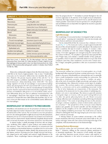
Peritoneal macrophages Mucosa-associated lymphoid tissue On the stained blood film the monocyte has a diameter of 12 to
15 μm (Fig. 67–1). The monocyte nucleus occupies approximately half
Inflammatory tissues Gastrointestinal tract
the area of the cell and usually is eccentrically placed. The nucleus most
Epithelioid cells Genitourinary tract often is reniform, but may be round or irregular. It contains a char-
Exudate macrophages Endocrine organs acteristic chromatin net with fine strands bridging small chromatin
clumps. Chromatin aggregates are arranged along the internal side of
Multinucleate giant cells Central nervous system (microglia)
the nuclear membrane. The cytoplasm is spread out, stains grayish-blue
Skin (histiocyte/dendritic cells) with Wright stain, and contains a variable number of fine, pink-pur-
ple granules, which at times are sufficiently numerous to give the entire
Data from Lewis, C, McGee, JD: The Macrophage, 2nd ed., Oxford cytoplasm a pink hue. Clear cytoplasmic vacuoles and a variable num-
University Press, New York, NY, 1992; Gordon S, Fraser I, Nath D. et al:
Macrophages in tissues and in vitro. Curr Opin Immunol 4:25-32, 1992; ber of larger azurophilic granulations often are encountered in these
Lasser A: The mononuclear phagocytic system: A review. Hum Pathol cells.
14:108-26, 1983.
Phase Microscopy
Monocytes continuously emigrate from the blood into tissue, with The monocyte nucleus has a distinct chromatin pattern on a cloudy
a half-life in the blood of approximately 1 day in mice. Nondividing background when examined by phase-contrast microscopy. The cyto-
6
monocytes can be induced to differentiate into dendritic like cells in plasm is clear gray. Mitochondria are extremely fine and occasionally
vitro. However, this process requires culture of the cells for 7 to 10 form a small, juxtanuclear rosette surrounding the centrosome. The
days with exogenous cytokines, typically interleukin (IL)-4 and gran- phase-dense cytoplasmic granules, varying in number, are gener-
7
ulocyte-monocyte colony-stimulating factor (GM-CSF). The major ally at the limit of resolution of light microscopy and appear as fine
lineage regulator of nearly all macrophages is monocyte/macrophage intracytoplasmic dust. Monocytes contain several types of cytoplas-
colony-stimulating factor (M-CSF; also termed CSF-1) and its receptor mic vacuoles. The reniform nucleus with a juxtanuclear depression
(M-CSF R). The M-CSF R is a class III transmembrane tyrosine kinase filled by a centrosome and its active undulating movement similar
8
receptor, which is expressed on most mononuclear phagocytes. In the to that of other leukocytes are characteristic of the monocyte. The
presence of endothelial cells grown on an extracellular matrix, mono- locomotion of the monocyte has the same pattern of undulating cyto-
cytes differentiate along two distinct pathways: toward dendritic cells plasmic veils seen in macrophages. The monocyte generally assumes
or macrophages. Monocytes that migrate across endothelium in an a triangular shape as it moves, with one point trailing behind and
abluminal to luminal direction differentiate into dendritic cells. In con- the other two points advancing before the cell. Blood monocytes
trast, monocytes that remain in the subendothelial matrix differentiate undergo adherence and cytoplasmic spreading following attachment
14
into macrophages. to glass surfaces. The extent of spreading increases in the presence
of antigen–antibody complexes, certain divalent metals, and proteo-
lytic enzymes. 14,15 The spread form of the monocyte reveals that the
MORPHOLOGY OF MONOCYTE PRECURSORS nucleus and granules are located centrally and the abundant hya-
Monoblasts and promonocytes are the precursors of monocytes, bear- loplasm is in the periphery of the cell, terminating in a fringed bor-
ing finely dispersed nuclear chromatin and nucleoli when observed in der that displays undulating movement. The small monocyte may be
the stained film of the marrow. The monoblast is a very-low-prevalence difficult to distinguish from the large lymphocyte when examined by
marrow cell, indistinguishable by light microscopy from the myeloblast. phase-contrast microscopy.
Promonocytes are 12 to 18 μm in diameter (as measured on dried blood A striking feature on phase-contrast microscopy is the ruffled
films) and have characteristic deeply indented, irregularly shaped nuclei plasma membrane that forms prominent phase-dense folds at the cell
with condensed chromatin, and numerous cytoplasmic microfilaments. surface and edges. Some cells have a dense thickening at the edge of the
In animal studies, a small percentage of marrow cells are phago- cytoplasm, with microextensions on the thickened edge.
cytic, synthesize DNA, adhere to glass surfaces, and contain nonspe-
cific esterases. These cells have been referred to as promonocytes and Scanning Electron Microscopy
9
are considered as intermediate between monoblasts and the monocytes The monocyte surface has very prominent ruffles and small surface
of the blood. Cytochemical studies identify the promonocyte in nor- blebs. 16,17 Extensive ruffling on the monocyte plasma membrane is
9
mal human marrow. Promonocytes have deeply indented and irregu- of functional significance. The monocyte is both motile and phago-
larly shaped nuclei and bundled and scattered single filaments in the cytic, and these functions require physical contact with particles or
cytoplasm. These morphologic features distinguish the promonocyte cell surfaces. Reduction in the radius of curvature of the cell surface
Kaushansky_chapter 67_p1043-1074.indd 1046 9/21/15 10:42 AM