
Page 1076 - Williams Hematology ( PDFDrive )
P. 1076
1050 Part VIII: Monocytes and Macrophages Chapter 67: Structure, Receptors, and Functions of Monocytes and Macrophages 1051
A B
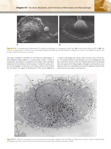
Figure 67–6. Scanning electron micrograph of cultured macrophages on coverglasses coated with (A) bovine serum albumin (BSA) or (B) with
immune complexes (BSA–anti-BSA). The macrophage develops prominent peripheral membrane ruffling and numerous microadhesion points to the
surface coated with immune complexes.
and rough endoplasmic reticulum are seen about the cell periphery. A is related to the phagocytic activity of the cell and its rate of pinocyto-
well-developed Golgi complex is in a juxtanuclear location. It often is sis. The number and size of mitochondria vary with the phagocytic and
multicentric and contains a concentration of vesicles, some with dense hence metabolic activity of the cell. Mitochondria tend to be grouped
inclusions that mark them as early lysosomes. A relatively constant fea- about the region of the Golgi complex, although several usually are seen
ture of cells engaged in endocytosis is the large number of microvilli at dispersed about the cell periphery, presumably supplying energy for the
the cell surface. The degree of development of this surface adaptation active endocytic processes occurring there.
N N
G G
Figure 67–7. Electron micrograph of monocyte-derived macrophage cultured in vitro for 9 days. G, Golgi zone; N, nucleus. Arrow on right indicates
endoplasmic reticulum; arrow on left indicates mitochondria; open arrow indicates lysosomes (×7600).
Kaushansky_chapter 67_p1043-1074.indd 1051 9/21/15 10:42 AM