
Page 1072 - Williams Hematology ( PDFDrive )
P. 1072
1046 Part VIII: Monocytes and Macrophages Chapter 67: Structure, Receptors, and Functions of Monocytes and Macrophages 1047
A B
C D
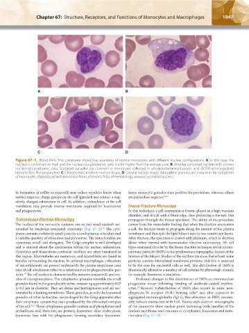
Figure 67–1. Blood films. This composite shows four examples of normal monocytes with different nuclear configurations. A. In this case, the
nucleus is contorted on itself and the nuclear-to-cytoplasmic ratio is a bit higher than the average case. B. Another contorted nucleus with a lower
nuclear-to-cytoplasmic ratio. Scattered vacuoles are common in monocytes collected in ethylenediaminetetraacetic acid (EDTA)-anticoagulated
blood before film preparation. C. Characteristic reniform nuclear shape. D. Circular nuclear shape. Azurophilic granules are evident in the cytoplasm
of monocytes. (Reproduced with permission from Lichtman’s Atlas of Hematology, www.accessmedicine.com.)
by formation of ruffles or microvilli may reduce repulsive forces when Some monocyte granules stain positive for peroxidase, whereas others
surface negative-charge groups on the cell approach and contact a neg- are peroxidase negative. 10,11
atively charged substratum or cell. In addition, redundancy of the cell
membrane may provide reserve membrane required for locomotion Freeze-Fracture Microscopy
and phagocytosis. In this technique, a cell suspension is frozen, placed in a high-vacuum
chamber, and struck with a blunt edge, thus producing a fracture that
Transmission Electron Microscopy propagates through the frozen specimen. The utility of the procedure
The nucleus of the monocyte contains one or two small nucleoli sur- comes from the remarkable finding that when the fracture encounters
rounded by nucleolar-associated chromatin (Fig. 67–2). The cyto- a cell, the fracture tends to propagate along the interior of the plasma
18
plasm contains a relatively small quantity of endoplasmic reticulum and membrane and thus split the lipid bilayer into its two constituent layers.
a variable quantity of ribosomes and polysomes. The mitochondria are After fracture, the specimen is coated with platinum, which is electron
numerous, small, and elongated. The Golgi complex is well developed dense when viewed with transmission electron microscopy. All cell
and is situated about the centrosome within the nuclear indentation. types examined thus far by the freeze-fracture technique reveal intram-
Centrioles and filamentous centriolar satellites are often visualized in embrane particles (IMPs) as the predominant topographic feature of the
this region. Microtubules are numerous, and microfibrils are found in interior of the bilayer. Studies of the erythrocyte show that at least some
bundles surrounding the nucleus. In cultured macrophages, collections particles contain intercalated membrane proteins, and this is assumed
of microfilaments are present underneath the plasma membrane near to be the case for nucleated cells as well. The distribution of IMPs is
sites of cell attachment either to a substratum or to phagocytosable par- dramatically altered in a number of cell systems by physiologic stimuli,
ticles. The cell surface is characterized by numerous microvilli and ves- for example, hormonal stimulation.
19
icles of micropinocytosis. The cytoplasmic granules resemble the small Profound changes in the distribution of IMPs on mononuclear
granules found in the granulocytic series, measuring approximately 0.05 phagocytes occur following binding of antibody-coated erythro-
to 0.2 μm in diameter. They are dense and homogeneous and are sur- cytes. Because redistribution of IMPs also occurs in some non-
13
rounded by a limiting membrane. These granules, as with the lysosomal phagocyte Fc receptor (FcR)–bearing cells and after exposure to
13
granules of other leukocytes, are packaged by the Golgi apparatus after aggregated immunoglobulin (Ig) G, this alteration in IMPs presum-
their enzymatic content has been produced by the ribosomal complex ably reflects interaction with FcR. Freeze-etch electron micrographs
of the cell. 10,11 These cytoplasmic granules contain acid phosphatase and of the monocyte show nuclear pores traversing both lamellae of the
arylsulfatase and, therefore, are primary lysosomes. After endocytosis, nuclear membrane and contours of cytoplasmic lysosomes and mito-
lysosomes fuse with the phagosome, forming secondary lysosomes. chondria (Fig. 67–3).
Kaushansky_chapter 67_p1043-1074.indd 1047 9/21/15 10:42 AM