
Page 125 - Williams Hematology ( PDFDrive )
P. 125
100 Part III: Epochal Hematology Chapter 7: Hematology of the Fetus and Newborn 101
28
dose–response in vitro to G-CSF. G-CSF is expressed by hepatocytes at the brain, are ultimately derived from the yolk sac of the embryo and
14 weeks’ gestation. 29 undergo limited maintenance after birth from hematopoietic stem
cells. 49,50
Lymphopoiesis
Lymphopoiesis is present in the lymph plexuses and the thymus begin- SYNTHESIS OF FETAL HEMOGLOBINS
20
ning at 9 weeks’ gestation. B cells with surface immunoglobulin (Ig) M
are present in the liver, and circulating lymphocytes also are seen at 9 Human hemoglobin (Hgb) is a tetramer composed of two α-type and
weeks’ gestation. T lymphocytes are found only rarely before 12 weeks’ two β-type globin chains (Table 7–1). The α-globin gene cluster is
gestation. Lymphocyte subpopulations are detected by 13 weeks’ ges- located on chromosome 16 and contains the ζ gene 5′ to the pair of
30
31
tation in fetal liver. Absolute numbers of major lymphoid subsets in α-globin genes. The β-globin gene cluster is located on chromosome 11
A
G
51
20- to 26-week-old fetuses, as defined by the antigens CD2, CD3, CD4, and contains five globin genes oriented 5′ to 3′ as ε-γ -γ -δ-β. During
CD8, CD16, CD19, and CD20, are similar to those in newborns (see embryogenesis the genes on both chromosomes are activated sequen-
“Neonatal Lymphopoiesis” below). 32,33 tially from the 5′ to the 3′ end. This globin “switching” is related not
only to the relative positions of the globin genes within their respective
Marrow Hematopoiesis chromosomal clusters, but also to interacting upstream “locus control
Hematopoietic cells are first seen in the marrow of the 10- to 11-week regions.” 52
embryo, and they remain confined to the diaphyseal regions of long Hgb Gower-1 (ζ ε ) is the major hemoglobin in embryos younger
1
2 2
bones until 15 weeks’ gestation. Initially, there are approximately equal than 5 weeks’ gestation (see Table 7–1). Hgb Gower-2 (α ε ) has been
34
53
2 2
numbers of myeloid and erythroid cells in the fetal marrow. However, found in embryos with a gestational age as young as 4 weeks and is
myeloid cells predominate by 12 weeks’ gestation, and the myeloid-to- absent in embryos older than 13 weeks. Hgb Portland (ζ γ ) is found in
54
2 2
erythroid ratio approaches the adult level of 3:1 by 21 weeks’ gestation. young embryos, but persists in infants with homozygous α-thalassemia
20
Macrophage cells in the fetal marrow, but not in the fetal liver, express (Chap. 48). Synthesis of the ζ and ε chains decreases as those of the α
29
the lipopolysaccharide receptor CD14. The marrow becomes the and γ chains increase (Fig. 7–2). The ζ-to-α-globin switch precedes the
major site of hematopoiesis after the 24th week of gestation and remains ε-to-γ-globin switch as the liver replaces the yolk sac as the main site of
so throughout the remainder of fetal life. erythropoiesis. 9,55
Hgb F (α γ ) is the major hemoglobin of fetal life (see Fig. 7–2).
56
2 2
ONTOGENY OF HEMATOPOIETIC STEM CELLS Synthesis of Hgb A can be demonstrated in fetuses as young as 9 weeks’
57
The reconstitution of the entire hematopoietic system by transplantation gestation. In fetuses of 9 to 21 weeks’ gestation, the amount of Hgb A
(α β ) rises from 4 to 13 percent of the total hemoglobin. These levels
57
with cord blood indicates that hematopoietic stem cells are circulating of Hgb A have enabled the antenatal diagnosis of β-thalassemia using
2 2
35
in the blood at birth. The immunologic reconstitution of an immuno- globin-chain synthesis. After 34 to 36 weeks’ gestation the percentage of
deficient human fetus with fetal-liver-derived cells also indicates that Hgb A rises, whereas that of Hgb F decreases (see Fig. 7–2). The mean
36
hematopoietic stem cells exist in the late gestation fetal liver. It was synthesis of Hgb F in term infants was 59.0 ± 10 percent (1 SD) of total
first postulated that hematopoietic stem cells originate independently hemoglobin synthesis as assessed by C-leucine uptake. The amount
14
58
in each hematopoietic site (yolk sac, liver, and marrow) of the embryo. of Hgb F in blood varies in term infants from 53 to 95 percent of total
37
However, experiments in the mammalian embryo indicate that the hemoglobin. 59
liver rudiment, like the marrow, is seeded by exogenous hematopoi- The fetal hemoglobin concentration in blood decreases after birth
etic cells. 38,39 It was initially thought that the liver, and eventually the by approximately 3 percent per week and is generally less than 2 to 3
marrow, were seeded by yolk sac–derived hematopoietic stem cells. percent of the total hemoglobin by 6 months of age. This rate of decrease
40
However, experiments in avian and amphibian embryos indicate that in Hgb F production is closely related to the gestational age of the infant
the hematopoietic stem cells that ultimately provide for long-term adult and is not affected by the changes in environment and oxygen tension
hematopoiesis arise within the body of the embryo proper rather than that occur at the time of birth. Hgb A (α δ ) has not been detected in
60
from the yolk sac. 41,42 Subsequent investigations in the mouse embryo fetuses. Normal adult levels of Hgb A are achieved by 4 months of age.
2
2 2
61
indicate that stem cells capable of engrafting myeloablated adult recip- Increased proportions of Hgb F at birth have been reported in infants
2
ients originate in the aorta-gonad-mesonephros (AGM) region of the who are small for gestational age, who have experienced chronic intra-
43
embryo proper. Cells capable of long-term engraftment of immunode- uterine hypoxia, who have trisomy 13, or who have died from sudden
ficient mice also first originate at 35 days of gestation in the aorta region infant death syndrome (SIDS). 62–66 Decreased levels of Hgb F at birth are
of human embryos. This correlates anatomically with the transient found in trisomy 21. 67
44
appearance of clusters of CD34+ blood cells closely associated with the
ventral wall of the aorta in several mammalian species, including the
5 weeks’ gestation human embryo. 45,46 These findings, as well as direct
47
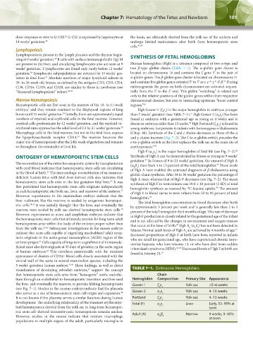
visualization of developing zebrafish embryos, support the concept TABLE 7–1. Embryonic Hemoglobins
that hematopoietic stem cells arise from “hemogenic” aortic endothe- Chain
lium through an endothelial-to-hematopoietic transition and then seed Hemoglobin Composition Primary Site Appearance
the liver, and eventually the marrow, to provide lifelong hematopoiesis Gower-1 ζ ε Yolk sac <5–6 weeks
(see Fig. 7–1). Studies in the murine embryo indicate that the placenta 2 2
48
also serves as a site of hematopoietic stem cell origin and expansion. Gower-2 α ε Yolk sac 4–13 weeks
2 2
It is not known if the placenta serves a similar function during human Portland ζ γ Yolk sac 4–13 weeks
2 2
development. The underlying relationship of the transient erythromye- Fetal (F) α γ Liver Early, 53–95% at
loid hematopoiesis derived from the yolk sac to long-term hematopoi- 2 2 term
etic stem cell–derived intraembryonic hematopoiesis remains unclear. Adult (A) α β Marrow 9 weeks, 5–45%
However, studies in the mouse indicate that resident macrophage 2 2 at term
populations in multiple organs of the adult, particularly microglia in
Kaushansky_chapter 07_p0097-0118.indd 101 9/18/15 10:13 PM