
Page 126 - Williams Hematology ( PDFDrive )
P. 126
102 Part III: Epochal Hematology Chapter 7: Hematology of the Fetus and Newborn 103
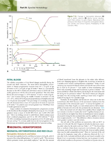
Figure 7–2. Changes in hemoglobin tetramers (A)
and in globin subunits (B) during human develop-
ment from embryo to early infancy. (Reproduced with
permission from Bunn HF, Forget BG: Hemoglobin: Molec-
ular, Genetic and Clinical Aspects. Philadelphia, PA: WB
Saunders; 1986.)
FETAL BLOOD of blood transferred from the placenta to the infant after delivery.
The cellular composition of fetal blood changes markedly during the Early cord clamping appears to heighten the occurrence of anemia at
75
second and third trimesters. The mean blood hemoglobin concen- 2 months and to impair cardiopulmonary adaptation. Delay of cord
tration in fetuses progressively increases from 9.0 ± 2.8 g/dL at age clamping may increase the blood volume and red cell mass of the infant
76,77
10 weeks to 16.5 ± 4.0 g/dL at age 39 weeks. There is a concomitant by as much as 55 percent. This results in fewer transfusions and
68
75
decrease in the MCV of fetal red cells from a mean of 134 fL/cell at 18 fewer days requiring oxygen and ventilation in preterm infants. The
weeks’ gestation to 118 fL/cell at 30 weeks’ gestation. The total white mean total blood volume after birth is 86 mL/kg for the term infant and
69
78
blood cell count averages 2 × 10 /L between 10 and 17 weeks of gesta- 89 mL/kg for the premature infant. The blood volume per kilogram
9
tion, and increases during the middle trimester to between 4.0 and decreases over the ensuing weeks, reaching a mean value of approxi-
31
4.5 × 10 /L, with an 80 to 85 percent preponderance of lymphocytes and mately 65 mL/kg by 3 to 4 months of age.
9
5 to 10 percent neutrophils. The percentage of circulating nucleated Normally the hemoglobin and hematocrit values rise in the first
69
red cells decreases from a mean of 12 percent at 18 weeks to 4 percent several hours after birth because of the movement of plasma from the
79
at 30 weeks. The platelet count remains greater than 150,000/μL from intravascular to the extravascular space. A venous hemoglobin con-
69
15 weeks’ gestation to term. 69,70 centration of less than 14 g/dL in a term infant and/or a fall in hemo-
Large numbers of committed hematopoietic progenitors circulate globin or hematocrit level in the first postnatal day are abnormal. Table
in the fetal blood. Blood samples obtained by fetoscopy at 12 to 19 weeks’ 7–2 shows the normal red cell values from capillary blood samples for
80
gestation reveal a mean of 20,450 BFU-E/mL and 12,490 CFU-GM/ term infants in the first 12 weeks after birth. Capillary hematocrit val-
mL. This is in striking contrast to adult blood, which contains many ues in newborns are higher than those in simultaneous venous samples,
71
fewer erythroid progenitors and 30 to 250 CFU-GM/mL. 70,72 Most particularly during the first postnatal days, and the capillary-to-venous
81
(70–80%) circulating hematopoietic progenitors at 26 to 28 weeks’ gesta- ratio is approximately 1.1:1. This difference reflects circulatory factors
tion are cycling. In contrast, adult-marrow-derived progenitors in the and is greater in preterm and sick infants.
72
bloodstream are relatively quiescent with only 0 to 5 percent cycling. The red cells of the newborn are macrocytic, with an MCV in
excess of 110 fL/cell. The MCV begins to fall after the first week, reach-
NEONATAL HEMATOPOIESIS ing adult values by the ninth week (see Table 7–2). 80,82 The blood film
from a newborn infant shows macrocytic normochromic cells, poly-
NEONATAL ERYTHROPOIESIS AND RED CELLS chromasia, and a few nucleated red blood cells. Even in healthy infants
83
there may be mild anisopoikilocytosis. Three to 5 percent of the red
Hemoglobin, Hematocrit, and Indices cells may be fragments, target cells, or otherwise distorted. By 3 to 5
The mean hemoglobin level in cord blood at term is 16.8 g/dL, with 95 days after birth, nucleated red blood cells are not found normally in the
73
percent of the values falling between 13.7 and 20.1 g/dL. This variation blood of term or premature infants, but they may be present in mark-
74
reflects perinatal events, particularly asphyxia, and also the amount edly elevated numbers in the presence of hemolysis or hypoxic stress. As
Kaushansky_chapter 07_p0097-0118.indd 102 9/18/15 10:13 PM