
Page 1525 - Williams Hematology ( PDFDrive )
P. 1525
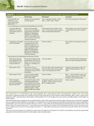
1500           Part XI:  Malignant Lymphoid Diseases                                                                                                        Chapter 90:  Classification of Malignant Lymphoid Disorders          1501


                TABLE 90–1.  Classification of Lymphoma and Lymphoid Leukemia by World Health Organization (Continued)
                Neoplasm               Morphology             Phenotype*                    Genotype †
                   Extranodal NK-cell   Angiocentric and angiode-  CD2+, cytoplasmic CD3ε +, CD4-,   No TCR rearrangement, EBV present
                  lymphoma, nasal-     structive growth       CD5-/+, CD7+, CD8-, CD56+
                  type (“angiocentric
                  lymphoma”) 1,45,46
                  Immunodeficiency-associated lymphoproliferative disorders
                   Lymphoproliferative   Range of morphology   Immunophenotype similar to that   FAS mutation seen in ALPS. Mutations
                    disorders associated   from reactive hyperplasia,   seen in immune competent patients   in SAP/SLAM in XLP. ATM mutations
                    with primary immune   polymorphous lymphoid   with corresponding malignancy  in AT
                    disorders          infiltrate, to high-grade
                                       lymphomas. Lymphoma
                                       and HL morphology is sim-
                                       ilar to that seen in immune
                                       competent patients
                   Lymphomas associated  Similar to above. Typical   Similar to above       MYC and BCL2 translocations seen in
                    with HIV infection  histologic features seen                            DLBCL
                                       in Burkitt lymphoma, HL,
                                       DLBCL. Lymphomas seen
                                       more frequently in HIV
                                       setting include primary
                                       effusion, plasmablastic,
                                       lymphomas, multicentric
                                       Castleman disease
                  Posttransplant lymphoproliferative disorders (PTLDs)
                     Early lesions: plasma-  PH: numerous plasma cells,  Similar to above   EBV+ in both IM and PH. Oligoclonal
                    cytic hyperplasia (PH)   lymphocytes, and immuno-                       polyclonal IgH rearrangement. EBV+
                    and infectious mono-  blasts. IM: numerous immu-
                    nucleosis (IM)-like  noblasts on a background
                                       of T cells
                   Polymorphic PTLD    Effacement of tissue   Similar to above with exception that   Clonal IG rearrangement. EBV+ by
                                       architecture with infiltrate   R-S cells in HLs often express CD30+,   EBER ISH. Mutated IgH in 75% of
                                       showing full range of B-cell  CD20+ but frequently are CD15-  cases
                                       maturation
                   Monomorphic PTLD    Similar to DLBCL, Burkitt   Similar to above         EBV+/-. Clonal B cell or T cells. Cyto-
                                       lymphoma or plasmacy-                                genetics frequently with TP53, RAS
                                       toma morphology                                      mutations, BCL6 translocations
                   Other iatrogenic    Increased frequency of HL   HL-like show CD20+, CD30+, CD15-   Same as above
                    immunodeficiency-   and lymphoproliferation   or CD20-, CD30+, CD15+ staining.
                    associated lymphopro-  with Hodgkin-like features.   EBV is variably positive
                    liferative disorders  Histologic features can oth-
                                       erwise resemble the range
                                       of features seen in other
                                       immune deficiency-related
                                       LPDs
               FISH, fluorescence  in situ hybridization; IgR, immunoglobulin gene rearrangement; IgVH, immunoglobulin variable heavy chain; MCD,
               multicentric castleman’s disease; neg., negative; NF-κB, nuclear factor-κB; NK, natural killer; R-S, Reed-Sternberg; SMZL, splenic marginal zone
               lymphoma; STAT, signal transducer and activator of transcription; TCR, T-cell receptor. Also see acronyms and abbreviations at the beginning
               of this chapter.
               * The immunophenotype revealed by immunohistochemistry and/or flow cytometry of surface antigens that typically are found for neoplastic
               cells of a given disorder are listed. If a CD antigen is indicated, then most of the neoplastic cells express that particular surface protein that is
               expressed by most tumor cells. CD antigens that have a minus (-) sign suffix are characteristically not expressed by the neoplastic cells of that
               disease entity. CD antigens that have a +/- sign suffix are not expressed by the neoplastic cells of all patients with that entity or are expressed at
               low or variable levels on the tumor cells. Antigens that have a -/+ sign suffix are expressed at very low levels or by the tumor cells of a minority
               of patients.
               † The common genetic features associated with a given type of neoplasm are indicated. The numbers in parentheses provide the approximate
               proportion of cases that have the defined phenotype or genetic abnormality.


          Kaushansky_chapter 90_p1491-1504.indd   1500                                                                  9/21/15   4:07 PM