
Page 1526 - Williams Hematology ( PDFDrive )
P. 1526
1500 Part XI: Malignant Lymphoid Diseases Chapter 90: Classification of Malignant Lymphoid Disorders 1501
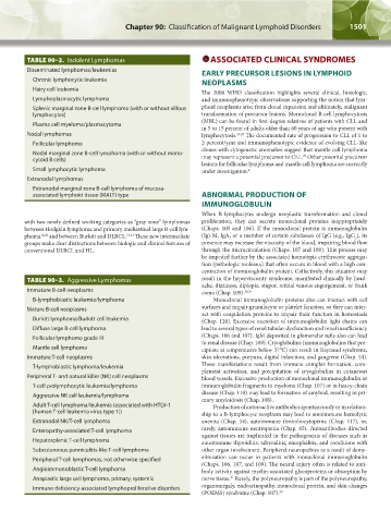
TABLE 90–2. Indolent Lymphomas ASSOCIATED CLINICAL SYNDROMES
Disseminated lymphomas/leukemias EARLY PRECURSOR LESIONS IN LYMPHOID
Chronic lymphocytic leukemia NEOPLASMS
Hairy cell leukemia
The 2008 WHO classification highlights several clinical, histologic,
Lymphoplasmacytic lymphoma and immunophenotypic observations supporting the notion that lym-
Splenic marginal zone B-cell lymphoma (with or without villous phoid neoplasms arise from clonal expansion and ultimately, malignant
lymphocytes) transformation of precursor lesions. Monoclonal B-cell lymphocytosis
(MBL) can be found in first-degree relatives of patients with CLL and
Plasma cell myeloma/plasmacytoma
in 5 to 15 percent of adults older than 60 years of age who present with
Nodal lymphomas lymphocytosis. 27,28 The documented rate of progression to CLL of 1 to
Follicular lymphoma 2 percent/year and immunophenotypic evidence of evolving CLL-like
clones with cytogenetic anomalies suggest that mantle cell lymphoma
Nodal marginal zone B-cell lymphoma (with or without mono- may represent a potential precursor to CLL. Other potential precursor
29
cytoid B cells)
lesions for follicular lymphoma and mantle cell lymphoma are currently
Small lymphocytic lymphoma under investigation. 4
Extranodal lymphomas
Extranodal marginal zone B-cell lymphoma of mucosa-
associated lymphoid tissue (MALT) type ABNORMAL PRODUCTION OF
IMMUNOGLOBULIN
When B lymphocytes undergo neoplastic transformation and clonal
with two newly defined working categories as “gray zone” lymphomas proliferation, they can secrete monoclonal proteins inappropriately
between Hodgkin lymphoma and primary mediastinal large B-cell lym- (Chaps. 105 and 106). If the monoclonal protein is immunoglobulin
phoma 12,26 and between Burkitt and DLBCL. 13,14 These new intermediate (Ig) M, IgA, or a member of certain subclasses of IgG (e.g., IgG ), its
3
groups make clear distinctions between biologic and clinical features of presence may increase the viscosity of the blood, impairing blood flow
conventional DLBCL and HL. through the microcirculation (Chaps. 107 and 109). This process may
be impeded further by the associated homotypic erythrocyte aggrega-
tion (pathologic rouleaux) that often occurs in blood with a high con-
centration of immunoglobulin protein. Collectively, this situation may
TABLE 90–3. Aggressive Lymphomas result in the hyperviscosity syndrome, manifested clinically by head-
ache, dizziness, diplopia, stupor, retinal venous engorgement, or frank
Immature B-cell neoplasms coma (Chap. 109). 30,31
B-lymphoblastic leukemia/lymphoma Monoclonal immunoglobulin proteins also can interact with cell
Mature B-cell neoplasms surfaces and impair granulocyte or platelet function, or they can inter-
act with coagulation proteins to impair their function in hemostasis
Burkitt lymphoma/Burkitt cell leukemia (Chap. 120). Excessive excretion of immunoglobulin light chains can
Diffuse large B-cell lymphoma lead to several types of renal tubular dysfunction and renal insufficiency
Follicular lymphoma grade III (Chaps. 106 and 107). IgM deposited in glomerular tufts also can lead
to renal disease (Chap. 109). Cryoglobulins (immunoglobulins that pre-
Mantle cell lymphoma cipitate at temperatures below 37°C) can result in Raynaud syndrome,
Immature T-cell neoplasms skin ulcerations, purpura, digital infarction, and gangrene (Chap. 54).
T-lymphoblastic lymphoma/leukemia These manifestations result from immune complex formation, com-
plement activation, and precipitation of cryoglobulins in cutaneous
Peripheral T- and natural killer (NK) cell neoplasms blood vessels. Excessive production of monoclonal immunoglobulin or
T-cell prolymphocytic leukemia/lymphoma immunoglobulin fragments in myeloma (Chap. 107) or in heavy-chain
disease (Chap. 110) may lead to formation of amyloid, resulting in pri-
Aggressive NK cell leukemia/lymphoma
mary amyloidosis (Chap. 108).
Adult T-cell lymphoma/leukemia (associated with HTLV-1 Production of autoreactive antibodies spontaneously or in relation-
[human T-cell leukemia virus type 1]) ship to a B-lymphocyte neoplasm may lead to autoimmune hemolytic
Extranodal NK/T-cell lymphoma anemia (Chap. 54), autoimmune thrombocytopenia (Chap. 117), or,
Enteropathy-associated T-cell lymphoma rarely, autoimmune neutropenia (Chap. 65). Autoantibodies directed
against tissues are implicated in the pathogenesis of diseases such as
Hepatosplenic T-cell lymphoma autoimmune thyroiditis, adrenalitis, encephalitis, and conditions with
Subcutaneous panniculitis-like T-cell lymphoma other organ involvement. Peripheral neuropathies as a result of demy-
Peripheral T-cell lymphomas, not otherwise specified elinization can occur in patients with monoclonal immunoglobulin
(Chaps. 106, 107, and 109). The neural injury often is related to anti-
Angioimmunoblastic T-cell lymphoma body activity against myelin-associated glycoproteins or absorption by
31
Anaplastic large cell lymphoma, primary, systemic nerve tissue. Rarely, the polyneuropathy is part of the polyneuropathy,
Immune deficiency-associated lymphoproliferative disorders organomegaly, endocrinopathy, monoclonal protein, and skin changes
(POEMS) syndrome (Chap. 107). 32
Kaushansky_chapter 90_p1491-1504.indd 1501 9/21/15 4:08 PM