
Page 1524 - Williams Hematology ( PDFDrive )
P. 1524
1498 Part XI: Malignant Lymphoid Diseases Chapter 90: Classification of Malignant Lymphoid Disorders 1499
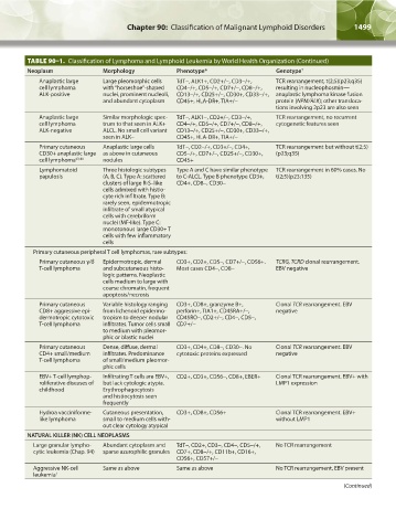
TABLE 90–1. Classification of Lymphoma and Lymphoid Leukemia by World Health Organization (Continued)
Neoplasm Morphology Phenotype* Genotype †
Anaplastic large Large pleomorphic cells TdT-, ALK1+, CD2+/-, CD3-/+, TCR rearrangement, t(2;5)(p23;q35)
cell lymphoma with “horseshoe”-shaped CD4-/+, CD5-/+, CD7+/-, CD8-/+, resulting in nucleophosmin—
ALK-positive nuclei, prominent nucleoli, CD13-/+, CD25+/-, CD30+, CD33-/+, anaplastic lymphoma kinase fusion
and abundant cytoplasm CD45+, HLA-DR+, TIA+/- protein (NPM/ALK); other transloca-
tions involving 2p23 are also seen
Anaplastic large Similar morphologic spec- TdT-, ALK1-, CD2+/-, CD3-/+, TCR rearrangement, no recurrent
cell lymphoma trum to that seen in ALK+ CD4-/+, CD5-/+, CD7+/-, CD8-/+, cytogenetic features seen
ALK-negative ALCL. No small cell variant CD13-/+, CD25+/-, CD30+, CD33-/+,
seen in ALK– CD45+, HLA-DR+, TIA+/-
Primary cutaneous Anaplastic large cells TdT-, CD2-/+, CD3+/-, CD4+, TCR rearrangement but without t(2;5)
CD30+ anaplastic large as above in cutaneous CD5-/+, CD7+/-, CD25+/-, CD30+, (p23;q35)
cell lymphoma 43,44 nodules CD45+
Lymphomatoid Three histologic subtypes Type A and C have similar phenotype TCR rearrangement in 60% cases. No
papulosis (A, B, C). Type A: scattered to C-ALCL. Type B phenotype CD3+, t(2;5)(p23;135)
clusters of large R-S–like CD4+, CD8-, CD30-
cells admixed with histio-
cyte rich infiltrate. Type B:
rarely seen, epidermotropic
infiltrate of small atypical
cells with cerebriform
nuclei (MF-like). Type C:
monotonous large CD30+ T
cells with few inflammatory
cells
Primary cutaneous peripheral T cell lymphomas, rare subtypes:
Primary cutaneous γ/δ Epidermotropic, dermal CD3+, CD2+, CD5-, CD7+/-, CD56+. TCRG, TCRD clonal rearrangement.
T-cell lymphoma and subcutaneous histo- Most cases CD4-, CD8- EBV negative
logic patterns. Neoplastic
cells medium to large with
coarse chromatin, frequent
apoptosis/necrosis
Primary cutaneous Variable histology ranging CD3+, CD8+, granzyme B+, Clonal TCR rearrangement. EBV
CD8+ aggressive epi- from lichenoid epidermo- perforin+, TIA1+, CD45RA+/-, negative
dermotropic cytotoxic tropism to deeper nodular CD45RO-, CD2+/-, CD4-, CD5-,
T-cell lymphoma infiltrates. Tumor cells small CD7+/-
to medium with pleomor-
phic or blastic nuclei
Primary cutaneous Dense, diffuse, dermal CD3+, CD4+, CD8-, CD30-. No Clonal TCR rearrangement. EBV
CD4+ small/medium infiltrates. Predominance cytotoxic proteins expressed negative
T-cell lymphoma of small/medium pleomor-
phic cells
EBV+ T-cell lymphop- Infiltrating T cells are EBV+, CD2+, CD3+, CD56-, CD8+, EBER+ Clonal TCR rearrangement. EBV+ with
roliferative diseases of but lack cytologic atypia. LMP1 expression
childhood Erythrophagocytosis
and histiocytosis seen
frequently
Hydroa vacciniforme- Cutaneous presentation, CD3+, CD8+, CD56+ Clonal TCR rearrangement. EBV+
like lymphoma small to medium cells with- without LMP1
out clear cytology atypical
NATURAL KILLER (NK) CELL NEOPLASMS
Large granular lympho- Abundant cytoplasm and TdT-, CD2+, CD3-, CD4-, CD5-/+, No TCR rearrangement
cytic leukemia (Chap. 94) sparse azurophilic granules CD7+, CD8-/+, CD11b+, CD16+,
CD56+, CD57+/-
Aggressive NK-cell Same as above Same as above No TCR rearrangement, EBV present
leukemia 1
(Continued)
Kaushansky_chapter 90_p1491-1504.indd 1499 9/21/15 4:07 PM