
Page 1521 - Williams Hematology ( PDFDrive )
P. 1521
1496 Part XI: Malignant Lymphoid Diseases Chapter 90: Classification of Malignant Lymphoid Disorders 1497
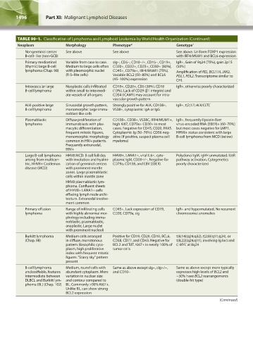
TABLE 90–1. Classification of Lymphoma and Lymphoid Leukemia by World Health Organization (Continued)
Neoplasm Morphology Phenotype* Genotype †
Nongerminal center See above See above See above. Uniform FOXP1 expression
B-cell–like (non-GCB) with IRF4/MUM1 and BCL6 expression
Primary mediastinal Variable from case to case. sIg-, CD5-, CD10-/+, CD15-, CD19+, IgR+, Gain of 9q24 (75%), gain 2p15
(thymic) large B-cell Medium to large cells often CD20+, CD22+, CD23+, CD30+ (80%), (50%)
lymphoma (Chap. 98) with pleomorphic nuclei CD45+, CD79a+, IRF4/MUM1 (75%). Amplification of REL, BCL11A, JAK2,
(R-S–like cells) Variable BCL2 (50–80%) and BCL6 PDL1, PDL2. Transcriptome similar to
(45–100%) expression CHL
Intravascular large Neoplastic cells infiltrated CD19+, CD20+, CD5 (38%), CD10 IgR+, otherwise poorly characterized
B-cell lymphoma within small to intermedi- (13%). Lack of CD29 (β1 integrin) and
ate vessels of all organs CD54 (ICAM1) may account for intra-
vascular growth pattern
ALK-positive large Sinusoidal growth pattern, Strongly positive for ALK, CD138+, IgR+, t(2;17) ALK/CLTC
B-cell lymphoma monomorphic large immu- VS38+, cytoplasmic IgA or IgG
noblast-like cells
Plasmablastic Diffuse proliferation of CD138+, CD38+, VS38C, IRF4/MUM1+, IgR+, frequently Epstein-Barr
lymphoma immunoblasts with plas- high Ki67, CD79a+ CD30+ in most virus-encoded RNA (EBER)+ (60–70%)
macytic differentiation, cases. Negative for CD45, CD20, PAX5. but most cases negative for LMP1.
frequent mitotic figures, Cytoplasmic Ig (50–70%). CD56 neg- HHV8+ status consistent with large
monomorphic morphology ative (if positive, suspect plasma cell B-cell lymphoma from MCD (below)
common in HIV+ patients. myeloma)
Frequently extranodal,
EBV+
Large B-cell lymphoma HHV8 MCD: B cell follicles HHV8+, LANA1+, viral IL6+, cyto- Polyclonal IgM. IgVH unmutated. IL6R
arising from multicen- with involution and hyalini- plasmic IgM, CD20+/-, Negative for pathway activation. Cytogenetics
tric, HHV8+ Castleman zation of germinal centers CD79a, CD138, and EBV (EBER) poorly characterized
disease (MCD) with prominent mantle
zones. Large plasmablastic
cells within mantle zone
HHV8 plasmablastic lym-
phoma. Confluent sheets
of HHV8+ LANA1+ cells
effacing lymph node archi-
tecture. Extranodal involve-
ment common
Primary effusion Range of infiltrating cells CD45+, Lack expression of CD19, IgR+ and hypermutated. No recurrent
lymphoma with highly abnormal mor- CD20, CD79a, sIg chromosomal anomalies
phology including immu-
noblastic, plasmablastic,
anaplastic. Large nuclei
with prominent nucleoli
Burkitt lymphoma Medium cells arranged Positive for CD19, CD20, CD10, BCL6, t(8;14)(q24;q32), t(2;8)(q11;q24), or
(Chap. 98) in diffuse, monotonous CD38, CD77, and CD43. Negative for t(8;22)(q24;q11), involving Ig loci and
pattern. Basophilic cyto- BCL2 and TdT. Ki67+ in nearly 100% of C-MYC at 8q24
plasm, high proliferative tumor cells
index with frequent mitotic
figures. “Starry sky” pattern
present
B-cell lymphoma Medium, round cells with Same as above except sIg-, cIg+/-, Same as above except more typically
unclassifiable, features abundant cytoplasm. More and CD10- expresses high levels of BCL2 and
intermediate between variation in nuclear size ~30% have BCL2 rearrangements
DLBCL and Burkitt lym- and contour compared to (double-hit type)
phoma (BL) (Chap. 102) BL. Commonly >90% Ki67+.
Unlike BL, can show strong
BCL2 expression
(Continued)
Kaushansky_chapter 90_p1491-1504.indd 1496 9/21/15 4:07 PM