
Page 1522 - Williams Hematology ( PDFDrive )
P. 1522
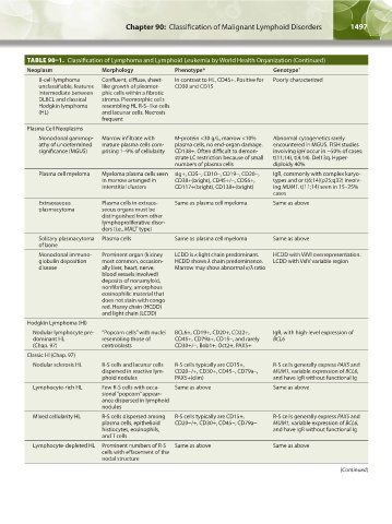
1496  Part XI:  Malignant Lymphoid Diseases     Chapter 90:  Classification of Malignant Lymphoid Disorders          1497


                   TABLE 90–1.  Classification of Lymphoma and Lymphoid Leukemia by World Health Organization (Continued)
                   Neoplasm               Morphology            Phenotype*                    Genotype †
                      B-cell lymphoma     Confluent, diffuse, sheet-  In contrast to HL, CD45+. Positive for   Poorly characterized
                      unclassifiable, features   like growth of pleomor-  CD30 and CD15
                      intermediate between   phic cells within a fibrotic
                      DLBCL and classical   stroma. Pleomorphic cells
                      Hodgkin lymphoma    resembling HL R-S–like cells
                      (HL)                and lacunar cells. Necrosis
                                          frequent
                   Plasma Cell Neoplasms
                      Monoclonal gammop-  Marrow infiltrate with   M-protein <30 g/L, marrow <10%   Abnormal cytogenetics rarely
                      athy of undetermined   mature plasma cells com-  plasma cells, no end-organ damage.   encountered in MGUS. FISH studies
                      significance (MGUS)  prising 1–9% of cellularity  CD138+. Often difficult to demon-  involving IgH occur in ~50% of cases:
                                                                strate LC restriction because of small   t(11;14), t(4;14). Del13q. Hyper-
                                                                numbers of plasma cells       diploidy 40%
                       Plasma cell myeloma  Myeloma plasma cells seen  sIg+, CD5-, CD10-, CD19-, CD20-,   IgR, commonly with complex karyo-
                                          in marrow arranged in   CD38+(bright), CD45+/-, CD56+,   types and or t(6;14)(p25;q32) involv-
                                          interstitial clusters  CD117+(bright), CD138+(bright)  ing MUM1. t(11;14) seen in 15–25%
                                                                                              cases
                      Extraosseous        Plasma cells in extraos-  Same as plasma cell myeloma  Same as above
                      plasmacytoma        seous organs must be
                                          distinguished from other
                                          lymphoproliferative disor-
                                          ders (i.e., MALT type)
                      Solitary plasmacytoma   Plasma cells      Same as plasma cell myeloma   Same as above
                      of bone
                      Monoclonal immuno-  Prominent organ (kidney   LCDD is κ light chain predominant.   HCDD with VλVI overrepresentation.
                      globulin deposition   most common, occasion-  HCDD shows λ chain predominance.   LCDD with VκIV variable region
                      disease             ally liver, heart, nerve,   Marrow may show abnormal κ/λ ratio
                                          blood vessels involved)
                                          deposits of nonamyloid,
                                          nonfibrillary, amorphous
                                          eosinophilic material that
                                          does not stain with congo
                                          red. Heavy chain (HCDD)
                                          and light chain (LCDD)
                   Hodgkin Lymphoma (Hl)
                      Nodular lymphocyte pre-  “Popcorn cells” with nuclei   BCL6+, CD19+, CD20+, CD22+,   IgR, with high-level expression of
                    dominant HL           resembling those of   CD45+, CD79a+, CD15-, and rarely   BCL6
                    (Chap. 97)            centroblasts          CD30+/-, Bob1+, Oct2+, PAX5+
                   Classic Hl (Chap. 97)
                     Nodular sclerosis HL  R-S cells and lacunar cells   R-S cells typically are CD15+,   R-S cells generally express PAX5 and
                                          dispersed in reactive lym-  CD20-/+, CD30+, CD45-, CD79a-,   MUM1, variable expression of BCL6,
                                          phoid nodules         PAX5+(dim)                    and have IgR without functional Ig
                     Lymphocyte-rich HL   Few R-S cells with occa-  Same as above             Same as above
                                          sional “popcorn” appear-
                                          ance dispersed in lymphoid
                                          nodules
                     Mixed cellularity HL  R-S cells dispersed among   R-S cells typically are CD15+,    R-S cells generally express PAX5 and
                                          plasma cells, epithelioid   CD20-/+, CD30+, CD45-, CD79a-  MUM1, variable expression of BCL6,
                                          histiocytes, eosinophils,                           and have IgR without functional Ig
                                          and T cells
                      Lymphocyte-depleted HL  Prominent numbers of R-S   Same as above        Same as above
                                          cells with effacement of the
                                          nodal structure
                                                                                                                   (Continued)


          Kaushansky_chapter 90_p1491-1504.indd   1497                                                                  9/21/15   4:07 PM