
Page 1531 - Williams Hematology ( PDFDrive )
P. 1531
1506 Part XI: Malignant Lymphoid Diseases Chapter 91: Acute Lymphoblastic Leukemia 1507
1 10
0.9 85.6% ± 2.9% XV (n = 498) 2000–2007 9 Male
0.8 78.9% ± 1.9% XIII–XIV (n = 465) 1991–1999 8 Total
0.7 70.6% ± 1.9% XI–XII (n = 546) 1984–1991 7 Female
Probability 0.5 53.5% ± 2.4% X (n = 428) 1979–1983 Rate per 100,000 6
0.6
5
0.4
0.3 35.5% ± 1.7% V–IX (n = 828) 1967–1979 4
0.2 3
0.1 8.9% ± 2.8% I–IV (n = 90) 1962–1966 2
0
0 5 10 15 20 25 30 35 40 45 1
Years from diagnosis 0
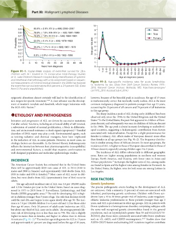
Figure 91–1. Kaplan-Meier analysis of event-free survival for 2855 <1 1–4 5–9 10–14 15–19 20–24 25–29 30–34 35–39 40–44 45–49 50–54 55–59 60–64 65–69 70–74 75–79 80–84 85+
children with ALL treated in 15 consecutive total-therapy studies
at St. Jude Children’s Research Hospital. Early intensification of systemic Age at diagnosis (years)
and intrathecal chemotherapy with a risk assignment based on sequen-
tial measurements of minimal residual disease in the 2000s has boosted Figure 91–2. Age-specific incidence rates for acute lymphoblas-
the event-free survival estimate to 85.6 percent ± 2.9 percent (SE). (Data tic leukemia by sex. (Data from SEER Cancer Statistics Review, 1975–
from CH Pui and is unpublished.) 2010, National Cancer Institute. Bethesda, MD, http://seer.cancer.gov/
csr/1975_2010. Accessed July 4, 2014.)
epigenetic alterations almost certainly will lead to the identification of however, because of the bimodal peak in incidence, the age of 13 years
new targets for specific treatment. 19,20 A clear advance was the develop- is mathematically correct but medically nearly useless. ALL is the most
ment of imatinib mesylate and dasatinib, which target leukemias with common malignancy diagnosed in patients younger than age 15 years,
the BCR-ABL1 fusion. 21 accounting for 23 percent of all cancers and 76 percent of all leukemias
in this age group.
The sharp incidence peak of ALL during early childhood has been
ETIOLOGY AND PATHOGENESIS observed only since the 1930s in the United Kingdom and the United
24
Initiation and progression of ALL are driven by successive mutations States. In the United States, the peak first appeared in children of Euro-
that alter cellular functions, including an enhanced ability of self-renewal, pean descent, and subsequently was seen in children of African descent
a subversion of control of normal proliferation, a block in differentia- in the 1960s. The age peak is absent in many developing or underdevel-
tion, and an increased resistance to death signals (apoptosis). Familial oped countries, suggesting a leukemogenic contribution from factors
1,2
disorders of DNA repair may play a role. Environmental agents, such associated with industrialization. Except for a slight predominance for
as ionizing radiation and chemical mutagens, have been implicated females in infancy, ALL affects males of European descent more often
in the induction of ALL in some patients. However, in most cases, no than females in all age groups (see Fig. 91–2). The frequency distribu-
etiologic factors are discernible. In the favored theory, leukemogenesis tion is similar among those of African descent. In most age groups, the
reflects the interaction between host pharmacogenetics (susceptibility) incidence of ALL is higher in those of European descent than in those of
and environmental factors, a model that requires confirmation in African descent, especially among children ages 2 to 3 years.
well-designed population and molecular epidemiologic studies. The incidence of ALL differs substantially in different geographic
areas. Rates are higher among populations in northern and western
INCIDENCE Europe, North America, and Oceania, with lower rates in Asian and
African populations. In Europe, the highest rates of ALL among males
25
The American Cancer Society has estimated that in the United States are found in Spain and the highest rates among females in Denmark. In
there will be approximately 6020 new cases of ALL in 2014 (3140 in the United States, the highest rates for both sexes are among Latinos in
males and 2880 in females) and approximately 1440 deaths from ALL Los Angeles.
22
(810 in males and 630 in females). Most cases of ALL occur in chil-
dren, but most deaths from ALL (approximately four of five) occur in RISK FACTORS
adults.
The age-adjusted incidence rate of ALL was 1.6 per 100,000 males Genetic Syndromes
and 1.2 for females per year in the United States, based on cases diag- The precise pathogenetic events leading to the development of ALL
nosed in 1975 to 2010 from 17 Surveillance, Epidemiology, and End are unknown. Only a minority (5 percent) of cases are associated with
Results (SEER) geographic areas. The risk for developing ALL is high- inherited, predisposing genetic syndromes. Children with Down syn-
23
est in children younger than 5 years of age. The risk then declines slowly drome have a 10 to 30 times greater risk of leukemia; acute megakary-
until the mid-20s, and begins to rise again slowly after age 50. The inci- oblastic leukemia predominates in those patients younger than age 3
dence is 7.9 per 100,000 children 1 to 4 years old and 1.2 for those older years, and ALL is predominant in older age groups. ALL in patients with
than age 60 years. Only 20 percent of adult acute leukemias are ALL, Down syndrome is a heterogeneous disorder, comprising subtypes with
but about one-third of ALL cases are in adults. The average person’s life- the same well-recognized genetic abnormalities found in the general
time risk of developing ALL is less than one in 750. The risk is slightly population, such as hyperdiploidy greater than 50 and t(12;21)[ETV6-
higher in males than in females, and higher in whites than in African RUNX1], plus those more commonly associated with Down syndrome
23
Americans (Fig. 91–2). The median age at diagnosis for ALL is 13 years such as +X, del(9), and CEBPD rearrangement. 26,27 Studies show that
and approximately 61 percent are diagnosed before the age of 20 years, P2RY8-CRLF2 fusion and activating JAK mutations together contribute
Kaushansky_chapter 91_p1505-1526.indd 1506 9/21/15 12:19 PM