
Page 1535 - Williams Hematology ( PDFDrive )
P. 1535
1510 Part XI: Malignant Lymphoid Diseases Chapter 91: Acute Lymphoblastic Leukemia 1511
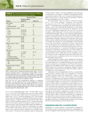
TABLE 91–3. Presenting Laboratory Features in Children 5 by the enhancer element of the immunoglobulin heavy-chain gene
on chromosome 14 is thought to contribute to leukemogenesis and the
and Adults with Acute Lymphoblastic Leukemia
72
associated eosinophilia in these cases. In patients with anemia, hemo-
Percent of Total globin levels are lower in younger children. Occasionally, a child with
Children, White/ ALL has a hemoglobin level as low as 1 g/dL.
Feature Black (%) Adults (%) Decreased platelet counts are common at diagnosis (median:
9
48–52 × 10 /L). Severe bleeding is uncommon, even when plate-
Cell lineage 9
T cell 15/24 25 let counts are as low as 20 × 10 /L, provided infection and fever are
73,74
B-cell precursor 85/76 75 absent. Occasional patients, principally male, present with throm-
bocytosis (>400 × 10 /L). Pancytopenia followed by a period of spon-
9
Leukocyte count (× 10 /L) taneous hematopoietic recovery may precede the diagnosis of ALL in
9
<10 47–49/34 41 rare cases. Coagulopathy, usually mild, can be seen in 3 to 5 percent
75
10–49 28–31/29 31 of patients, most of whom have T-cell ALL, and is only rarely associ-
50–99 8–12/14 12 ated with clinical bleeding. The level of serum lactate dehydrogenase is
76
>100 11–13/23 16 increased in most patients with ALL and is well correlated with tumor
Hemoglobin concentration (g/dL) burden. Increased levels of serum uric acid are common in patients with
<8 48/58 28 a large leukemia cell burden, reflecting an increased rate of purine cata-
8–10 24/22 26 bolism. Leukemic infiltration of the kidneys can lead to increased levels
>10 28/20 46 of creatinine, urea nitrogen, uric acid, and phosphorus. Rarely, patients
Platelet count (× 10 /L) present with hypercalcemia resulting from release of parathyroid
9
<50 46/40 52 hormone-like protein from lymphoblasts and leukemic infiltration of
50–100 23/20 22 bone. The t(17;19)(q22;p13.3) with E2A-HLF fusion, found in 0.5 per-
>100 31/40 26 cent of B-cell precursor ALL, is associated with adolescent age group,
77
disseminated coagulopathy, hypercalcemia, and dismal prognosis.
CNS status* Liver dysfunction as a result of leukemic infiltration occurs in 10 to 20
CNS1 67–79/60 92–95 percent of patients and is usually mild. However, recognition of carri-
CNS2 5–24/27 ? ers of hepatitis B virus is important because prompt lamivudine ther-
CNS3 3/3 5–8
Traumatic lumbar 6–7/10 ? apy can prevent serious complications from virus reactivation during
78
puncture with blasts immunosuppressive treatment.
Chest radiography is needed to detect enlargement of the thymus
Leukemic blasts in marrow (%) or mediastinal nodes and pleural effusions (see Fig. 91–3). Although
<90 33/46 29 bony abnormalities, such as metaphyseal banding, periosteal reactions,
>90 67/54 71 osteolysis, osteosclerosis, and osteopenia, can be found in 50 percent of
Leukemic blasts in blood patients, especially children with low leukocyte counts at presentation,
Present 87/90 92 skeletal films are not necessary for management. Magnetic resonance
Absent 13/10 8 imaging (MRI) is useful in patients with suspected vertebral collapse or
meningeal or nerve root involvement.
* CNS1, no blast cells in cerebrospinal fluid sample; CNS2, <5 leuko- Examination of the cerebrospinal fluid (CSF) is an essential diag-
cytes/μL with blast cells in a nontraumatic sample; CNS3, ≥5 leuko- nostic procedure. Leukemic blasts can be identified in as many as
cytes/μL with blast cells in a nontraumatic sample or the presence
of a cranial nerve palsy; and traumatic lumbar puncture with blasts one-third of pediatric patients and approximately 5 percent of adult
(≥10 erythrocytes/μL with blasts). Data on CNS2 and traumatic lum- patients at diagnosis of ALL; most of these patients lack neurologic
bar puncture with blasts are not available in adults. symptoms. 79,80 Traditionally, CNS leukemia is defined by the presence
Data from Pui CH: Acute lymphoblastic leukemia, in Childhood of at least 5 leukocytes/μL of CSF (with leukemic blast cells apparent in
Leukemias, 2nd ed, edited by CH Pui, p 439. Cambridge University a cytocentrifuged sample) or by the presence of cranial nerve palsies.
Press, New York, 2006 and Larson RA, Dodge RK, Burns CP, et al: A However, with the omission of prophylactic cranial irradiation in con-
five-drug remission induction regimen with intensive consolidation temporary clinical trials, the presence of any leukemic blast cells in the
for adults with acute lymphoblastic leukemia: Cancer and Leukemia CSF is associated with increased risk of CNS relapse and is an indica-
Group B study 8811. Blood 85:2025, 1995. tion to intensify intrathecal therapy. Different opinions exist regarding
80
when the first lumbar puncture should be performed. Most protocols
10–12 × 10 /L). Hyperleukocytosis (>100 × 10 white cells/L) occurs now require the procedure at diagnosis and instill the first dose of
9
9
in 11 to 13 percent of white children, but occurs more often in black chemotherapeutic agents intrathecally. Contamination of the CSF by
children (23 percent) and in adults (16 percent) because they are more leukemia cells as a result of traumatic lumbar puncture at diagnosis is
81
likely to have T-cell ALL. Profound neutropenia (<0.5 × 10 /L) is found associated with an inferior treatment outcome in children with ALL.
9
in 20 to 40 percent of patients, elevating their risk for infection. Approx- The risk of traumatic lumbar puncture can be decreased by administer-
imately 90 percent of patients have circulating leukemic blast cells at ing platelet transfusions to thrombocytopenic patients and by having
diagnosis. Hypereosinophilia, generally reactive, may precede the diag- the most experienced clinician perform the procedure.
nosis of ALL by several months. Some patients, mostly male, have ALL
with the t(5;14)(q31;q32) chromosomal abnormality and a hypereosin-
ophilic syndrome (pulmonary infiltration, cardiomegaly, and conges- DIAGNOSIS AND CELL CLASSIFICATION
tive heart failure). These patients often do not have circulating leukemic Examination of a marrow aspirate is recommended for diagnosis of
blasts or other cytopenias and have a relatively low percentage of blasts ALL because as many as 10 percent of patients lack circulating blasts at
in the marrow. Activation of the interleukin-3 gene on chromosome the time of diagnosis and because high concentrations of marrow cells
72
Kaushansky_chapter 91_p1505-1526.indd 1510 9/21/15 12:19 PM