
Page 1707 - Williams Hematology ( PDFDrive )
P. 1707
1682 Part XI: Malignant Lymphoid Diseases Chapter 103: Cutaneous T-Cell Lymphoma (Mycosis Fungoides and Sézary Syndrome) 1683
A B C
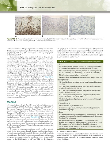
Figure 103–3. Mycoses fungoides. Immunohistochemistry. A. CD4+ lichenoid infiltrate in the superficial dermis. Note Pautrier microabscess in the
epidermis. B. Few CD8+ cells are present. C. Loss of maturation marker CD7.
with a predominance of larger atypical cells extending deeper into the tomography (CT) and positron emission tomography (PET) scans are
33
dermis; epidermotropism may be lost. Transformation to large T-cell used to assess involvement of lymph nodes. 50,51 Excisional lymph node
lymphoma (CD30+ or CD30−) may occur and carries a poor prognosis biopsy is usually recommended to assess the extent of the disease and
in the setting of MF. 34 nodal architecture, but other techniques, including fine-needle aspira-
Immunophenotyping plays an important role in diagnosis. The tion, are selectively used as well. 52
cells usually are CD3+CD4+CD45RO+CD8−, a phenotype associated
with mature helper-inducer T lymphocytes. Loss of maturation mark- TABLE 103–2. TNMB Classification of Mycosis Fungoides
ers, such as CD7 and CD26 expression on CD4+ are important markers
for malignant T lymphocytes. 35,36 The cells may express T-cell activa- T: Skin
tion markers, such as HLA-DR or CD25 (interleukin [IL]-2 receptor). T1: Limited patches, papules, or plaques covering <10% of the
Clonal rearrangement of the T-cell receptor (TCR) Vβ gene can be skin surface (T1a = patch only; T1b = plaques ± patches)
identified in approximately 90 percent of advanced cases of MF, but in T2: Generalized patches, papules, or plaques covering 10% of
37
only 50 percent of early stage cases. In rare instances, the classic clin- the skin surface (T2a = patch only; T2b = plaques ± patches)
ical presentation of MF may be associated with an aberrant CD4 phe- T3: At least one tumor (≥1 cm in diameter)
notype or may have CD4−dyCD8+ T-cell phenotype 38,39 (Fig. 103–3A T4: Generalized erythroderma over at least 80% body surface area
to C). Recent molecular studies have identified several new markers, N: Lymph nodes
which may prove useful as positive markers of the disease, including
thymocyte selection-associated high mobility group box factor N0: No clinically abnormal peripheral lymph nodes; biopsy not
(TOX), plastin (PLS3), and killer cell immunoglobulin-like receptor required
KIR3DL2. 40–43 Cytogenetic abnormalities are not consistently identi- N1: Clinically abnormal peripheral lymph nodes; histopathol-
fied, but loss of heterozygosity on 10q and microsatellite instability may ogy Dutch grade 1 or NCI LN0 to 2
44
be seen in advanced-stage disease. A possible association exists with N2: Clinically abnormal peripheral lymph nodes; histopathol-
homozygous deletion of PTEN and CDKN2A and tumor-suppressor ogy Dutch grade 2 or NCI LN3
genes on 10p and 9p chromosomes, respectively. These may be silenced N3: Clinically abnormal peripheral lymph nodes; histopathol-
with progression of disease. 45,46 ogy Dutch grades 3 to 4 or NCI LN4
NX: Clinically abnormal peripheral lymph nodes; no histologic
confirmation
STAGING M: Visceral organs
MF is classified according to the widely accepted modified tumor, node, M0: No visceral organ involvement
metastasis, blood (TNMB) classification, originally adopted in 1975 by
the Mycosis Fungoides Cooperative Study Group 47,48 and revised to M1: Visceral organ involvement; requires histologic confirma-
tion and specify organ
match modern developments in the field. Accurate determination of
16
the stage in MF and SS is of utmost importance because of its prognostic B: Blood
significance and critical role in selecting therapy. Cutaneous lesions are B0: Atypical circulating cells not present (<5%); specify “a” if flow
classified using the T staging system (Table 103–2). The area of the skin cytometry is negative for clonal T lymphocytes or “b” if positive
and type of the lesions were found to correlate with patient survival and for clonal T lymphocytes
are important prognostic predictors and need to be calculated at every B1: Atypical circulating cells present (>5%, minimal blood
visit to assess disease status. Prognosis varies according to tumor bur- involvement); specify “a” if flow cytometry is negative for clonal
49
den. The presence of tumors (T3) may indicate a worse prognosis than T lymphocytes or “b” if positive for clonal T lymphocytes
erythroderma (T4). 18 B2: Leukemia (≥1000 cells/μL, CD4 to CD8 ratio of 10 or higher,
The extent of extracutaneous disease usually correlates with the evidence of a T-cell clone in the blood)
extent of skin involvement. In early disease, significant involvement NCI, National Cancer Institute.
of lymph nodes and blood is unlikely. However, lymphadenopathy is T indicates the size of the tumor and whether it has invaded nearby
present in more than half of patients as disease advances and increases tissue. N indicates the regional lymph nodes that are involved. M
with progressive cutaneous involvement. Lymph nodes are assigned N indicates distant metastasis. B indicates whether there are tumor
category in the TNMB staging of MF (see Table 103–2). Computed cells in the blood.
16
Kaushansky_chapter 103_p1679-1692.indd 1682 9/21/15 12:50 PM