
Page 1705 - Williams Hematology ( PDFDrive )
P. 1705
1680 Part XI: Malignant Lymphoid Diseases Chapter 103: Cutaneous T-Cell Lymphoma (Mycosis Fungoides and Sézary Syndrome) 1681
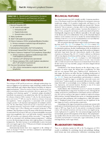
TABLE 103–1. World Health Organization–European CLINICAL FEATURES
Organization for Research and Treatment of Cancer The clinical presentation of MF is highly variable. Cutaneous manifesta-
Classification of Primary Cutaneous T-Cell and Natural tions of the disease result from skin infiltration by malignant cutaneous
Killer Cell Lymphomas lymphocyte antigen (CLA)-positive lymphocytes and depend on the
I. Mycosis Fungoides (MF) extent of skin involvement. Patients initially may present with “chronic
A. MF variants and subtypes dermatitis” that is resistant to therapy, which is often misdiagnosed as
1. Folliculotropic MF spongiotic dermatitis (so-called eczema), “psoriasis-like dermatitis,” or
other chronic, nonspecific dermatoses, usually associated with pruritus.
2. Pagetoid reticulosis Histologically, diagnosis may be difficult, especially in the early stages
3. Granulomatous slack skin of the disease and in its erythrodermic form, as the abnormal atypical
II. Sézary Syndrome infiltrate can be minimal and can be masked by normal inflammatory
III. Adult T-Cell Leukemia/Lymphoma infiltrates in the skin, or it can be misinterpreted as a normal inflamma-
IV. Primary Cutaneous CD30+ Lymphoproliferative Disorders tory infiltrate because of its mature CD4+ phenotype.
MF may progress through distinct stages of skin involvement, rang-
A. Primary cutaneous anaplastic large cell lymphoma ing from patch (Fig. 103–1A) to plaque (Fig. 103–1B) to tumor (Fig.
B. Lymphomatoid papulosis 103–1C), but any type of lesion may progress or lesions may arise de novo.
V. Subcutaneous Panniculitis-Like T-Cell Lymphoma For descriptive purposes, the skin manifestations of MF are divided into
VI. Extranodal Natural Killer/T-Cell Lymphoma, Nasal Type patch stage (patch-only disease), plaque stage (both patches and plaques),
VII. Primary Cutaneous Peripheral T-Cell Lymphoma, Unspecified and tumor stage (more than one tumor present, usually in the context
A. Primary cutaneous aggressive epidermotropic CD8+ of patches and plaques) (Fig. 103–1C). A patch is defined as a flat lesion
T-cell lymphoma (provisional) with various degrees of erythema and fine scaling; it may be atrophic or
B. Cutaneous γδ T-cell lymphoma (provisional) poikilodermatous (Fig. 103–1D). A plaque is a well-demarcated
erythematous, brownish, or violaceous lesion of at least 1 mm elevation
C. Primary cutaneous CD4+ small-/medium-size pleomor- with a variable amount of scale. Tumors are elevated at least 10 mm above
phic T-cell lymphoma (provisional) the skin surface and may resemble a plaque or be dome shaped without
VIII. Precursor Hematologic Neoplasm significant scaling.
A. CD4+/CD56+ hematodermic neoplasm (blastic NK-cell Distribution of the lesions depends on the clinical stage at pre-
lymphoma) sentation. In earlier stages, the lesions have a predilection for folds
and non–sun-exposed body areas (“bathing trunk” distribution). In
later stages, the lesions can affect the face, including development of
ectropion, and other areas, such as palms and soles (keratoderma; see
Fig. 103–1E). Tumors may be generalized, and ulceration is common.
ETIOLOGY AND PATHOGENESIS Progression through the stages is variable but commonly occurs over
several years. Lesions usually are associated with pruritus, which may
29
The etiologies of MF and SS are unknown, although epidemiologic fea- range from mild to excruciatingly severe, leading to insomnia, weight
tures are suggestive of an infectious origin, including a predilection for loss, depression, and suicidal ideation. Pruritus is one of the most
elderly individuals and a higher-than-expected incidence in immune- important quality-of-life issues for these patients. 30
suppressed patients. However, studies to date have failed to reveal con- Erythrodermic skin involvement occurs in 5 percent of patients
19
sistent associations between any particular infectious agent and CTCL, with MF. Manifestations range from very faint to severe, with significant
including novel infectious agents. A “persistent antigen stimulation” scaling, keratoderma, painful fissures of the hands and feet, nail dystro-
20
hypothesis was proposed as an initial event after MF was observed to phy, and nail loss leading to the patient’s inability to walk and maintain
be a disease of mature CD4+ memory T cells, but the stimulating anti- daily activities. Severely inflamed skin serves as a breeding ground for
gen is not known. 21,22 MF also may be viewed as a disease of immune bacteria and other pathogens, with resulting fevers, chills, and septice-
dysregulation. Tumor progression is associated with decreased mia. 19,31 Peripheral edema of the extremities may be significant in the
antigen-specific T-cell responses and impaired cell-mediated cytotox- later stages and lead to cardiovascular compromise.
icity. On the other hand, improved survival is associated with intact Depending on the stage of presentation, patients may present with
cell-mediated immunity. Progression of MF is associated with progres- nodal and/or blood involvement and/or visceral metastases. The earliest
sive T-helper type 2 (Th2) skewing and increased production of Th2 stages of MF (e.g., Stage IA and B), may pursue a waxing and waning
cytokines. This alteration accounts for many of the immune abnormali- course, with minimal symptoms and may not have any negative prog-
ties associated with advanced MF, such as hypereosinophilia, increased nostic implications for a patient. The more advanced stages usually, but
serum immunoglobulin (Ig) A and IgE levels, impaired natural killer not inevitably, present with symptoms of the disease. The symptomatol-
(NK) cell function, and impaired cellular immunity. 23,24 Late-stage MF ogy usually reflects the site and severity of involvement and ranges from
and SS are associated with declining immunocompetence, resulting completely asymptomatic to severe pain, organ malfunction, or at the
25
in severe life-threatening infections, and a high incidence of secondary end stage disease, multi-organ failure.
malignancies. The latter increase is not attributable to prior treatment
with carcinogenic agents alone. Fifty percent of deaths among patients LABORATORY FINDINGS
26
with MF result from infections.
Environmental factors have been suggested in the etiology of There is no definitive marker for MF or SS. The diagnosis of CTCL
CTCL in Europe, 27,28 but have not been confirmed by epidemiologic usually is established by correlating clinical and pathologic findings.
studies in the United States. Early lesions may show polymorphic infiltration (containing mixed
Kaushansky_chapter 103_p1679-1692.indd 1680 9/21/15 12:50 PM