
Page 1708 - Williams Hematology ( PDFDrive )
P. 1708
1682 Part XI: Malignant Lymphoid Diseases Chapter 103: Cutaneous T-Cell Lymphoma (Mycosis Fungoides and Sézary Syndrome) 1683
TABLE 103–3. Revised Staging of Mycosis Fungoides and as B category in the TNMB staging (see Table 103–2). For staging pur-
poses, the B2 blood rating is equivalent to nodal involvement.
The B2
58,59
Sézary Syndrome 16
rating is defined as (1) a Sézary cell count of 1000 cells/mm or more; (2)
3
T N M B a CD4-to-CD8 ratio of 10 or higher caused by an increase in circulat-
IA 1 0 0 0, 1 ing T cells and/or an aberrant loss or expression of pan–T-cell markers
by flow cytometry; (3) increased lymphocyte counts with evidence of a
IB 2 0 0 0, 1
T-cell clone in the blood determined by the Southern blot or PCR tech-
IIA 1, 2 1, 2 0 0, 1 nique; or (4) a chromosomally abnormal T-cell clone. Malignant cells
IIB 3 0–2 0 0, 1 also can be detected using sensitive techniques such as cytogenetics or
TCR gene rearrangement studies. 60–64 Patients with blood involvement
III 4 0–2 0 0, 1
have a higher likelihood of lymphadenopathy and visceral involvement.
IIIA 4 0–2 0 0 Marrow infiltration is infrequently detected by biopsy despite circu-
IIIB 4 0–2 0 1 lating malignant cells; it is identified at autopsy in 30 to 40 percent of
cases. The cytologic appearance of the malignant cells in visceral organs
IVA1 1–4 0–2 0 2
is similar to that of the malignant cells in the skin. 65
IVA2 1–4 3 0 0–2 In the erythrodermic subset of MF, three T4 subsets can be identi-
IVB 1–4 0–3 1 0–2 fied (Table 103–4). In general, in SS, a triad of exfoliative erythroderma,
generalized lymphadenopathy, and leukemia, has the worst prognosis
See Table 103–2 for definitions of T1 to T4, N0 to N3, and M0 to M1. among the forms of MF.
DIFFERENTIAL DIAGNOSIS
Histopathologic examination of affected lymph nodes may show
partial or complete effacement of normal architecture, with a monomor- Diagnosis of MF is based on a constellation of findings, which include
phic infiltrate of MF cells. However, in most cases, the nodal architec- clinical presentation, skin and lymph node biopsies (if indicated), and
ture is not effaced, and dermatopathic changes are present with varying blood evaluation. A number of benign dermatoses can mimic MF or SS,
numbers of atypical lymphocytes in the T-cell paracortical areas of the and may even have TCR gene rearrangements. 66–69 Such benign condi-
node. Even the presence of dermatopathic changes alone in the lymph tions include psoriasis and psoriasiform dermatoses (such as pityriasis
nodes carries prognostic significance (see Tables 103–2 and 103–3). 53,54 rubra pilaris, seborrheic dermatitis, contact dermatitis, and eczema),
Abnormal lymph nodes should be biopsied regardless of the T stage. intertrigo, tinea, drug eruptions, and other conditions.
Metastatic disease (including patients with positive nodes) is the Cutaneous and systemic lymphomas other than MF should be con-
most significant prognostic predictor (see Tables 103–2 and 103–3). sidered in the differential diagnosis. Smoldering adult T-cell leukemia/
Patients with visceral involvement that includes liver, spleen, pleura, lymphoma has a number of clinical features similar to MF, but it usually
55
and lung have a median survival of less than 1 year. Blood involvement can be distinguished by the presence of antibodies to human T-lympho-
may be an important predictor of progression and survival (Figs. 103–4 tropic virus type 1 (HTLV-1) and by other associated findings unusual
and 103–5). The number of circulating Sézary cells increases with in MF. However, this distinction may be difficult. 70,71
56
advancing disease, and the cells are particularly prominent in patients Pagetoid reticulosis (Woringer-Kolopp disease) is a rare skin dis-
with generalized erythroderma. However, even in early stage disease, a order that consists of solitary or localized cutaneous plaques. It affects
high frequency of clonal T cells in the blood may be detected using a young males almost exclusively. It has a benign course, and the prog-
highly sensitive polymerase chain reaction (PCR) technique, suggesting nosis is excellent. 72–74 It is an epidermal process, with the majority of
that early systemic disease is common. Blood involvement is rated atypical lymphocytes found within hyperplastic epidermis. Although
75
57
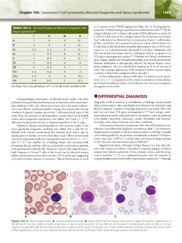
A B C
Figure 103–4. Blood lymphocytes. A. Normal small lymphocyte. B. Sézary cell. Note the nuclear swirls and the light microscopic appearance of
the Sézary cell nucleus. Without careful inspection in cases of lymphocytosis, Sézary cells can be mistaken for small lymphocytes as seen in chronic
lymphocytic leukemia. C. Blood lymphocytes from a patient with mycosis fungoides and disseminated disease involving marrow and blood. Note
clefted appearance of the nucleus. (Reproduced with permission from Lichtman’s Atlas of Hematology, www.accessmedicine.com.)
Kaushansky_chapter 103_p1679-1692.indd 1683 9/21/15 12:50 PM