
Page 1765 - Williams Hematology ( PDFDrive )
P. 1765
1740 Part XI: Malignant Lymphoid Diseases Chapter 107: Myeloma 1741
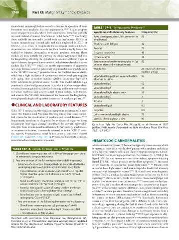
monoclonal immunoglobulins, osteolytic lesions, suppression of bone TABLE 107–3. Symptomatic Myeloma 247
formation near myeloma foci and angiogenesis. 224,225 Studies propose
novel xenogeneic models, where three-dimensional bone-like scaffolds Symptoms and Laboratory Features Frequency (%)
are used instead of human fetal bone or rabbit bone. 226,227 Specifically, Bone pain (spine, chest, less common in 58
these scaffolds are internally coated with murine/human BMSCs or long bones)
human mesenchymal stromal cells and then implanted in SCID or Weakness and fatigue 32
RAG2–/–γc–/– mice, to recapitulate the autologous marrow microen-
vironment in vivo. Myeloma cells are then loaded directly inside the Anemia 73
scaffold or injected intracardiac to mimic myeloma homing. These Elevated creatinine 48
models are more suitable for studying the microenvironment and also Hypercalcemia 28
for drug testing, affording the opportunity to evaluate different stages or
type of disease. Syngeneic tumor models include transplantable murine Serum monoclonal immunoglobulin (Ig) 82
models like 5T33, 228,229 which are mostly representative of aggressive peak on standard electrophoresis
late-stage disease, as well as genetically engineered mouse models, such Weight loss 24 (one-half of whom
as the Vk*MYC mouse, generated from the C57BL/6 mouse strain, had lost ≥9 kg)
230
which has a high incidence of spontaneous monoclonal gammopathy Monoclonal Ig peak on immunofixation 97
with aging, after activation-induced cytidine deaminase–dependent of serum or urine
MYC activation in germinal center B-cells. This model exhibits high Monoclonal IgG 52
penetrance, clonal malignant plasma cells, which produce isotype class-
switched immunoglobulins, a similar histology and immunophenotype Monoclonal IgA 21
to human myeloma, and delayed onset of renal failure, bone lesions, Monoclonal light chains only 16
and anemia. The Vk*MYC mouse model has been used for drug testing,
strongly paralleling the drug activity observed in myeloma patients. 230 Monoclonal IgD 2
Biclonal 2
CLINICAL AND LABORATORY FEATURES Monoclonal IgM 0.5
Table 107–1 summarizes the signs and symptoms associated with mye- Negative 6.5
loma. The International Myeloma Working Group has issued simpli- Urinary monoclonal light chains 75
fied criteria for the classification of myeloma and related disorders. 231,232 Marrow plasmacytosis >10% 90
Symptomatic myeloma is diagnosed by evidence of organ or tissue
impairment (end-organ damage) manifested by anemia, hypercalce- Data from Kyle RA, Gertz MA, Witzig TE, et al: Review of 1027
mia, lytic bone lesions, renal insufficiency, hyperviscosity, amyloidosis, patients with newly diagnosed multiple myeloma. Mayo Clin Proc
or recurrent infections, (commonly referred to as the “CRAB” crite- 78:21–33, 2003.
ria; namely, hypercalcemia, renal failure, anemia, and bone lesions)
(Tables 107–2 and 107–3; Fig. 107–6). Presence of these features neces-
sitate immediate treatment for myeloma. HEMATOLOGIC ABNORMALITIES
Myelomatous involvement of the marrow typically causes anemia, which
TABLE 107–2. Criteria for Diagnosis of Myeloma is present in more than two-thirds of patients with myeloma and relates
to the degree of marrow infiltration. The erythropoietin response is insuf-
1. Clonal bone marrow plasma cells ≥10% of biopsy-proven bony ficient in myeloma, owing to production of cytokines, (IL-1, TNF-β, Fas
or extramedullary plasmacytoma ligand, MIP-1α, and tumor necrosis factor– related apoptosis-inducing
2. Any one or more of the following myeloma-defining events: ligand [TRAIL], which produce erythroblast apoptosis), increased
233
• Evidence of end-organ damage that can be attributed to the serum viscosity, or concomitant renal dysfunction. 234–236 Patients with
underlying plasma cell proliferative disorder, specifically: myeloma have high urinary and serum hepcidin levels that inversely
• Hypercalcemia: serum calcium >0.25 mmol/L (>1 mg/dL) correlate with hemoglobin values. 237,238 IL-6 and bone morphogenetic
higher than the upper limit of normal or >2.75 mmol/L protein (BMP)-2 mediate hepcidin transcription in the liver via STAT3
(>11 mg/dL) signaling, which, in turn, blocks iron release from macrophages and
239
• Renal insufficiency: creatinine clearance <40 mL per min or inhibits iron absorption from the intestine. 240,241 In contrast to other lym-
serum creatinine >177 µmol/L (>2 mg/dL) phoproliferative disorders, thrombocytopenia is uncommon at diagno-
• Anemia: hemoglobin value of >20 g/L below the lower sis, even with extensive marrow infiltration, as IL-6 has thrombopoietic
limit of normal, or a hemoglobin value <100 g/L activity. In some patients, thrombocytopenia might occur secondary
242
• Bone lesions: one or more osteolytic lesions on skeletal to treatment or to autoimmune mechanisms (such as those accounting
Δ
radiography, CT, or PET-CT for anemia or factor VIII deficiency 46,243–245 ). Specifically, bortezomib
• Any one or more of the following biomarkers of malignancy: causes a cyclic thrombocytopenia, with a different kinetic from cyto-
• Clonal bone marrow plasma cell percentage* ≥60% toxic drugs, appearing during the first 10 days of each cycle, but with
• Involved: uninvolved serum free light chain ratio ≥100 a short recovery time, no cumulative or persistent effects and absence
of marrow megakaryocyte toxic damage, as it primarily results from a
• >1 focal lesions on MRI studies functional alteration in platelet budding. Prolonged exposure to alky-
246
Modified with permission from Rajkumar SV, Dimopoulos MA, lating agents can also promote onset of a concomitant myelodysplastic
Palumbo A, et al: International Myeloma Working Group updated syndrome. Overt bleeding is a relatively uncommon presenting symp-
247
criteria for the diagnosis of multiple myeloma. Lancet Oncol 2014 tom for myeloma patients ; however it occurs more commonly with
Nov;15(12):e538–e54. IgA paraproteins, in the presence of very high concentrations of serum
Kaushansky_chapter 107_p1733-1772.indd 1740 9/21/15 12:34 PM