
Page 1800 - Williams Hematology ( PDFDrive )
P. 1800
1774 Part XI: Malignant Lymphoid Diseases Chapter 108: Immunoglobulin Light-Chain Amyloidosis 1775
TABLE 108–2. Amyloid Syndromes Seen at the Mayo
Clinic
% Present
Kidney 67
Heart 47
Peripheral nerve 12
Liver 12
Autonomic nerve 4
Carpal tunnel 12
Tongue 9
Table 108–2 gives the frequency of amyloid syndromes seen in
patients at the Mayo Clinic. If an oncologist sees a patient with one
of these five syndromes, or an internist is faced with an undiagnosed
patient, laboratory evaluation is indicated, as outlined below.
LABORATORY FEATURES
The best screening tests for initial evaluation of patients with suspected
amyloidosis are immunoelectrophoresis and immunofixation of both
serum and urine and a serum immunoglobulin free light-chain assay
(both κ and λ). Systemic immunoglobulin AL amyloidosis is a plasma
cell dyscrasia, and 99 percent of patients will have a detectable abnor-
mality of one of these three tests, reflecting synthesis by a clonal pop-
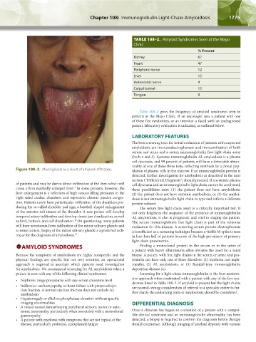
Figure 108–2. Macroglossia as a result of amyloid infiltration. ulation of plasma cells in the marrow. If an immunoglobulin protein is
detected, further investigation for amyloidosis as described in the next
section (“Differential Diagnosis”) should proceed. If a systemic plasma
of patients and may be due to direct infiltration of the liver, which will cell dyscrasia and an immunoglobulin light chain cannot be confirmed,
cause a firm markedly enlarged liver. In some patients, however, the three possibilities exist: (1) the patient does not have amyloidosis,
17
liver enlargement is a reflection of high venous filling pressures in the (2) the patient does not have systemic amyloidosis, or (3) the amyloi-
right-sided cardiac chambers and represents chronic passive conges- dosis is not immunoglobulin light chain in type and reflects a different
tion. Patients rarely have periarticular infiltration of the shoulders pro- protein subunit.
ducing the so-called shoulder pad sign, a baseball-shaped enlargement The serum free light-chain assay is a critically important test. It
of the anterior soft tissues of the shoulder. A rare patient will develop not only heightens the suspicion of the presence of immunoglobulin
temporal artery infiltration and develop classic jaw claudication, as well AL amyloidosis, it also is prognostic and vital to staging the patient.
as limb, buttock, and calf claudication. On questioning, many patients The serum immunoglobulin free light chain is part of the response
18
will have xerostomia from infiltration of the minor salivary glands; and evaluation for this disease. A screening serum protein electrophoresis
at some centers, biopsy of the minor salivary glands is a preferred tech- is insufficient as a screening technique because a visible M-spike is seen
nique for the diagnosis of amyloidosis. 19 in less than half of patients because of the high prevalence of primary
light-chain proteinemia.
AMYLOID SYNDROMES Finding a monoclonal protein in the serum or in the urine of
a patient with heavy albuminuria often obviates the need for a renal
Because the symptoms of amyloidosis are highly nonspecific and the biopsy. A patient with free light chains in the serum or urine and pro-
physical findings are specific but not very sensitive, an operational teinuria can have only one of three disorders: (1) myeloma cast neph-
approach is required to ascertain which patients need investigation ropathy, (2) AL amyloidosis, or (3) Randall-type immunoglobulin
for amyloidosis. We recommend screening for AL amyloidosis when a deposition disease (κ).
patient is seen with any of the following clinical syndromes: Screening for a light-chain immunoglobulin is the best noninva-
• Nephrotic range proteinuria with any serum creatinine level sive approach when confronted with a patient with any of the five syn-
• Infiltrative cardiomyopathy or heart failure with preserved ejec- dromes listed in Table 108–2. If amyloid is present but the light chains
are normal, strong consideration of referral to a specialty center to fur-
tion fraction. A normal ejection fraction does not exclude AL
amyloidosis ther clarify the underlying form of amyloidosis should be considered.
• Hepatomegaly or alkaline phosphatase elevation without specific
imaging abnormalities DIFFERENTIAL DIAGNOSIS
• A mixed axonal demyelinating peripheral sensory, motor or auto-
nomic neuropathy, particularly when associated with a monoclonal Once a clinician has begun an evaluation of a patient with a compat-
gammopathy ible clinical syndrome and an immunoglobulin abnormality has been
• A patient with myeloma with symptoms that are not typical of the detected, a biopsy is required to confirm the diagnosis before therapy
disease, particularly profound, unexplained fatigue should commence. Although imaging of amyloid deposits with various
Kaushansky_chapter 108_p1773-1784.indd 1775 9/18/15 9:53 AM