
Page 1803 - Williams Hematology ( PDFDrive )
P. 1803
1778 Part XI: Malignant Lymphoid Diseases Chapter 108: Immunoglobulin Light-Chain Amyloidosis 1779
A B
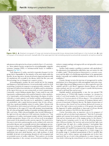
Figure 108–5. A. Polarized micrograph of Congo red–stained endomyocardial biopsy demonstrates birefringence in the involved area. B. Light
micrograph of hematoxylin-and-eosin–stained slide of the same specimen demonstrating amyloid as extracellular eosinophilic amorphous material.
subcutaneous fat aspiration has a lower sensitivity than in AL amyloido- ischemic symptomatology and angina with normal epicardial coronary
sis. These patients require recognition by echocardiography, magnetic artery anatomy. 40
resonance imaging (MRI), or endomyocardial biopsy to establish a Sudden death remains a problem in patients with amyloidosis.
41
diagnosis (Fig. 108-5). The placement of an implantable defibrillator may not reduce the risk
Early diagnosis of cardiac amyloid is imperative because it is this of sudden death. Electromechanical dissociation is a common occur-
42
group that is responsible for the majority of the early deaths with this rence and the ability of a fibrillating amyloid heart to be appropriately
disorder. In our experience, 40 percent of newly diagnosed patients with shocked internally and establish hemodynamic stability has not been
AL amyloidosis will succumb to the disease within the first year; and established. 43
this percentage has not changed over a quarter century. Diuretic therapy remains the mainstay of management for cardiac
Echocardiography remains the most useful test for the imaging amyloidosis; but in these patients with noncompliant ventricular cham-
and diagnosis of amyloid; however, it is not particularly useful for serial bers, higher than normal filling pressures are often required to open the
monitoring of patients for response or progression after treatment. The ventricle and fill it with blood. Aggressive diuretic therapy will often
technique still suffers from interobserver variability, and the calculations reduce preload, and this can result in drops in systolic blood pressure,
of the septal thickness can vary substantially on serial measurements. reduced renal blood flow, and syncope. 44
Conversely, a septal thickness greater than 15 mm would be rare with Familial amyloid cardiomyopathy is rare, but one special TTR
hypertensive cardiomyopathy and would be limited to either amyloido- mutant known as TTRVal122Ile requires awareness. In a prospective
sis or hypertrophic cardiomyopathy. Interestingly, the degree of cardiac study of cord blood samples, this mutation was found in 3 percent of
infiltration in senile systemic and familial amyloid cardiomyopathy is newborns of American parents of African descent. This compared with
substantially greater than that seen in AL amyloidosis. A patient with a prevalence of 0.44 percent in Americans of European descent and 0
AL amyloidosis with a septal thickness greater than 18 mm will gen- percent of Americans of Hispanic descent. The degree of penetrance of
erally have significant disability related to cardiac failure. Patients with this mutation at the clinical level has not been determined; however, in
TTR amyloidosis, both mutant and wild-type, will frequently have sep- view of the high incidence and prevalence of this genetic abnormality,
tal thicknesses in the range of 25 mm with minimal symptomatology. the diagnosis of cardiac amyloidosis in Americans of African descent
The old echocardiographic finding of granular sparkling appearance is warrants early analysis of the DNA for this mutation. 45,46
little used today. Other echocardiographic clues include thickening of Cardiac biomarkers play an important role in the prognosis of
the right ventricle and reduction in left ventricular chamber size. Late amyloid as well as in its functional assessment. Both the B-natriuretic
37
47
consequences of cardiac involvement include valvular thickening and peptide and troponin levels predict outcomes in patients with amyloi-
valvular regurgitation. Repair of the valve will not result in meaningful dosis and are important parts of the staging system for this disease.
48
improvement in the patient’s aerobic exercise capacity. The restriction to Staging involves assigning a point for any of the following character-
flow seen in restrictive cardiomyopathy can be confused with restrictive istics: difference between the involved and uninvolved free light chain
pericardial disease; and occasionally, patients have undergone pericar- of greater than 180 mg/L; for a cardiac troponin T level greater than
diectomies without benefit. Endomyocardial biopsy is highly sensitive 0.025 ng/mL; and for the N-terminal of the prohormone brain natri-
38
in the diagnosis of cardiac amyloidosis; and if five specimens are taken, uretic peptide (NT-proBNP) level greater than 1800 pg/mL. This creates
recognition of the diagnosis is virtually certain. Patients with cardiac four stages with median survivals ranging from 6 months (stage 4) to
amyloidosis have poor atrial function and a high incidence of atrial 60 months (stage 1). Serialized measurements of the NT-proBNP also
standstill. Atrial and atrial appendage thrombi are well recognized and have been used to define response and progression and, in fact, have
are potential sources of cardiac embolism. Rare patients can develop supplanted the use of serial echocardiography to assess changes over
39
amyloid deposition in the coronary microcirculation resulting in true time, both in following the natural history as well as assessing response
Kaushansky_chapter 108_p1773-1784.indd 1778 9/18/15 9:53 AM