
Page 1801 - Williams Hematology ( PDFDrive )
P. 1801
1776 Part XI: Malignant Lymphoid Diseases Chapter 108: Immunoglobulin Light-Chain Amyloidosis 1777
A B C
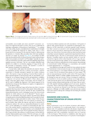
Figure 108–3. Technique and results of subcutaneous fat aspiration. A. Procedural technique. B. Fat stained with Congo red, note the preserved
interstices of the fat cells. C. Viewed under polarized light to demonstrate green birefringence.
radionuclides, most notably anti–serum amyloid P component, con- less than two-thirds of patients seen with amyloidosis. A full quarter of
tinue to be explored and used in practice, they are not a substitute for patients with amyloid deposits are composed of amyloidogenic tran-
histologic confirmation of the presence of amyloidosis. 20–22 In a patient sthyretin (ATTR) (inherited) or wild-type amyloid (senile systemic).
with renal, cardiac, hepatic, or peripheral nerve amyloidosis, it is straight Less than 4 percent are amyloid A (AA) and approximately 4 percent
forward to establish the diagnosis by kidney, heart, liver, or nerve represent ALect2 amyloidosis, which deposits in the kidney and causes
biopsy, but this is unnecessary in the majority of patients. Subcutaneous proteinuria. Other forms of amyloid, which are often localized, are seen
fat aspiration is a bedside office procedure done with local anesthesia in less than 1 percent of patients. A particularly important form of local-
and does not require an incision (Fig. 108–3). An excellent YouTube ized amyloid occurs at the sites of subcutaneous insulin injections in
video has been produced by the Boston Medical Center on the technique diabetics. Crystalline insulin can form amyloid, which can cause dis-
(https://www.youtube.com/watch?v=tctYTmxd9gQ). Typically, fat aspira- colored firm deposits which, when biopsied, will be Congo red–posi-
tions are reviewed at a specialty center unless the pathology department tive, but by mass spectroscopic analysis, can be confirmed to be insulin.
regularly processes fatty tissue. The fat is not paraffin-embedded and, Other forms of systemic amyloidosis for which systemic chemotherapy
therefore, it is subject to overfixation, trapping of Congo red dye, and is contraindicated include fibrinogen A-α amyloidosis, and amyloid
the possibility of inadequate controls. Fat aspiration is positive in caused by apolipoprotein-A1, and gelsolin.
23
three-quarters of patients with amyloidosis. One additional advantage, particularly for those patients who have
A second tissue easily stained for amyloid is the marrow. Because TTR amyloidosis is that mass spectroscopy not only identifies the pro-
AL amyloidosis patients will have a plasma cell dyscrasia, a marrow tein, in most instances, it can confirm whether the protein is wild-type
examination is clinically indicated to quantify the number of plasma or a mutant. We still recommend that patients who have mutant TTR
cells in the marrow. Congo red staining of marrow blood vessels can seen on mass spectroscopy have this confirmed by DNA sequencing of
detect amyloid deposits in half of patients with AL amyloidosis. Com- a blood sample, which is a readily available commercial test, to confirm
bining the two techniques (marrow and fat pad aspiration), establishes the findings on mass spectroscopy. We have investigated a small num-
a diagnosis in 85 percent of afflicted patients. Minor salivary gland ber of patients in whom an amyloid syndrome was strongly suspected,
biopsy, endoscopic gastric biopsy, rectal biopsy, and skin biopsy can a subcutaneous fat aspirate was not definitively positive, yet mass spec-
also be used to establish the diagnosis. If the clinical suspicion of AL troscopic analysis demonstrated peptides in the tissue that are associ-
amyloidosis is strong and these biopsies are negative, it is appropriate to ated with amyloid including apolipoprotein-E and serum amyloid P
biopsy the affected organ. component. When a pathologist makes a diagnosis of amyloid in tissue
Once tissue containing Congo red has been identified, it becomes sections, the clinicians are required to ask what type of amyloidosis it
imperative that the protein subunit be determined. Historically, immu- represents. Although not widely available, mass spectroscopic analysis
nohistochemistry has been used to identify the type of amyloid, but is the most sensitive and specific technique for identifying the amyloid
immunohistochemistry can be challenging because only those protein subunit protein.
subunits for which antisera exist can be detected. 24,25 Second, partic-
ularly in AL amyloidosis, the reactive epitope may have been deleted
during the process of amyloid fibril formation and, frequently, the back- DIAGNOSIS AND CLINICAL
ground staining is high, particularly in kidney tissues. Third, because CHARACTERIZATION OF ORGAN-SPECIFIC
the protein in amyloid misfolds, even if the epitopes are present, they
may be hidden deep within the deposits and may be inaccessible to SYNDROMES
commercial antisera. Not only can immunohistochemistry be unreli- Kidney
able, it can be misleading. It is therefore recommended that laser cap- Kidney involvement is seen in 45 percent of patients with immuno-
ture of the amyloid deposit be performed routinely followed by mass globulin AL amyloidosis. It is conspicuously absent in the majority of
spectroscopic analysis. 26–28 The subunit protein can be identified by this patients with TTR amyloidosis until late in the disease. Renal involve-
approach in the majority of cases. Even in patients who have a false-pos- ment is virtually universal in systemic AA amyloidosis as well as ALect2
itive Congo red stain, this technique is useful because it will not identify amyloidosis and A-fib α amyloidosis. Amyloid is seen in 2.5 percent
amyloid-related peptides upon analysis. With this technique, one is able of all renal biopsies. When limited to nondiabetics older than age 50
to confirm the presence of AL amyloidosis, which represents slightly years, amyloid deposits will be found in 10 percent of renal biopsies.
29
Kaushansky_chapter 108_p1773-1784.indd 1776 9/18/15 9:53 AM