
Page 1813 - Williams Hematology ( PDFDrive )
P. 1813
1788 Part XI: Malignant Lymphoid Diseases Chapter 109: Macroglobulinemia 1789
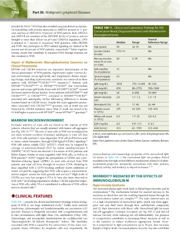
revealed by WGS. WGS has also revealed common defects in chroma- TABLE 109–1. Clinical and Laboratory Findings for 356
29
tin remodeling with somatic mutations in ARID1A present in 17 per-
cent, and loss of ARID1B in 70 percent of WM patients. Both ARID1A Consecutive Newly Diagnosed Patients with Waldenström
and ARID1B are members of the SWI/SNF family of proteins, and are Macroglobulinemia
thought to exert their effects via p53 and CDKN1A regulation. TP53 Normal
is mutated in 7 percent of sequenced WM genomes, while PRDM2 Median Range Reference Range
and TOP1 that participate in TP53-related signaling are deleted in 80 Age (years) 58 32–91 NA
29
percent and 60 percent of WM patients, respectively. Taken together,
somatic events that contribute to impaired DNA damage response are Gender (male/ 215/141 NA
female)
also common in WM.
Marrow involve- 30 5–95 NA
Impact of Waldenström Macroglobulinemia Genomics on ment (% of area
Clinical Presentation on slide)
MYD88 and CXCR4 mutations are important determinants of the Adenopathy 15 NA
clinical presentation of WM patients. Significantly higher marrow dis- (% of patients)
ease involvement, serum IgM levels, and symptomatic disease requir- Splenomegaly 10 NA
ing therapy, including hyperviscosity syndrome was observed in those (% of patients)
50
patients with MYD88 L265P CXCR4 WHIM/NS mutations. Patients with IgM (mg/dL) 2620 270–12,400 40–230
MYD88 L265P CXCR4 WHIM/FS or MYD88 L265P CXCR4 had intermediate
WT
WT
WT
marrow and serum IgM levels; those with MYD88 CXCR4 showed IgG (mg/dL) 674 80–2770 700–1600
the lowest marrow disease burden. Fewer patients with MYD88 L265P and IgA (mg/dL) 58 6–438 70–400
CXCR4 WHIM/FS or CXCR4 WHIM/NS , compared to MYD88 L265P CXCR4 Serum viscosity 2.0 1.1–7.2 1.4–1.9
WT
presented with adenopathy, further delineating differences in disease (cp)
tropism based on CXCR4 status. Despite the more-aggressive presen-
tation associated with CXCR4 WHIM/NS genotype, risk of death was not Hematocrit (%) 35 17–45 35–44
impacted by CXCR4 mutation status. Risk of death was found to be Platelet count 275 42–675 155–410
10-fold higher in patients with MYD88 versus MYD88 L265P genotype. 50 (× 10 /L)
9
WT
White cell count 6.4 1.7–22 3.8–9.2
9
MARROW MICROENVIRONMENT (× 10 /L)
Increased numbers of mast cells are found in the marrow of WM β M (mg/dL) 2.5 0.9–13.7 0–2.7
2
patients, wherein they are usually admixed with tumor cell aggregates LDH (U/mL) 313 61–1701 313–618
(see Fig. 109–1). 14,18,57 The role of mast cells in WM was investigated in
one study wherein coculture of primary autologous or mast cell lines β M, β -microglobulin; cp, centipoise; LDH, lactic dehydrogenase; NA,
2
2
with WM cells resulted in dose-dependent WM cell proliferation and/ not applicable.
or tumor colony formation, through CD40 ligand (CD40L) signaling. Data from patients seen at the Dana Farber Cancer Institute, Boston,
57
WM cells release soluble CD27 (sCD27), which may be triggered by MA.
cleavage of membrane-bound CD27 by matrix metalloproteinase 8
58
(MMP8). sCD27 levels are elevated in the serum of WM patients, and
follow disease burden in mice engrafted with WM cells, as well as in physicochemical and immunologic properties of the monoclonal IgM.
60
WM patients. sCD27 triggers the upregulation of CD40L and a pro- As shown in Table 109–2, the monoclonal IgM can produce clinical
liferation-inducing ligand (APRIL) on mast cells derived from WM manifestations through several different mechanisms related to its phys-
patients, and mast cell lines through its receptor CD70. Modeling in icochemical properties, nonspecific interactions with other proteins,
mice engrafted with a CD70-blocking antibody shows inhibition of antibody activity, and tendency to deposit in tissues. 61–63
tumor cell growth, suggesting that WM cells require a microenviron-
59
mental support system for their growth and survival. High levels of
CXCR4 and very late antigen-4 (VLA-4) have also been observed in MORBIDITY MEDIATED BY THE EFFECTS OF
WM cells. In blocking experiments studies, CXCR4 supported migra- IMMUNOGLOBULIN M
60
tion of WM cells, while VLA-4 contributed to adhesion of WM cells to Hyperviscosity Syndrome
marrow stromal cells. 60 The increased plasma IgM levels leads to blood hyperviscosity and its
complications. The mechanisms behind the marked increase in the
64
CLINICAL FEATURES resistance to blood flow and the resulting impaired transit through the
microcirculatory system are complex. 64–67 The main determinants are
Table 109–1 presents the clinical and laboratory findings at time of diag- (1) a high concentration of monoclonal IgMs, which may form aggre-
16
nosis of WM in one large institutional study. Unlike most indolent gates and may bind water through their carbohydrate component;
lymphomas, splenomegaly and lymphadenopathy are uncommon (≤15 and (2) their interaction with blood cells. Monoclonal IgM increases
percent). Purpura is frequently associated with cryoglobulinemia and red cell aggregation (rouleaux formation; see Fig. 109–1) and red cell
in rare circumstances with light-chain (AL) amyloidosis (Chap. 108). internal viscosity while reducing red cell deformability. The presence
Hemorrhagic and neuropathic manifestations are multifactorial (see of cryoglobulins contributes to increasing blood viscosity, as well as
“Immunoglobulin M–Related Neuropathy” below). The morbidity to the tendency to induce erythrocyte aggregation. Serum viscos-
associated with WM is caused by the concurrence of two main com- ity is proportional to IgM concentration up to 30 g/L, then increases
ponents: tissue infiltration by neoplastic cells and, importantly, the sharply at higher levels. Increased plasma viscosity may also contribute
Kaushansky_chapter 109_p1785-1802.indd 1788 9/21/15 12:30 PM