
Page 1817 - Williams Hematology ( PDFDrive )
P. 1817
1792 Part XI: Malignant Lymphoid Diseases Chapter 109: Macroglobulinemia 1793
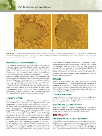
A B
Figure 109–5. Marrow clot section. A. Tryptase-staining mast cells surrounding a nodule of lymphoplasmacytic cells in a patient with Waldenström
macroglobulinemia. B. Mast cells in the same section exhibit strong CD40 ligand signaling, which has been shown to support (at least in part) the
growth and survival of lymphoplasmacytic cells.
IMMUNOLOGIC ABNORMALITIES scleral depression. In more severe cases of hyperviscosity, dot, blot,
68
and flame-shaped hemorrhages can appear in the macular area along
High-resolution electrophoresis combined with immunofixation of with markedly dilated and tortuous veins with focal constrictions
serum and urine is recommended for identification and characteriza- resulting in “venous sausaging,” as well as papilledema. The effects of
tion of the IgM monoclonal protein. The light chain of the monoclonal hyperviscosity are mediated by the blood viscosity as a result of IgM-
IgM is κ in 75 to 80 percent of patients. More than one M component red cell interactions but measurement of serum viscosity is much sim-
may be present. The concentration of the serum monoclonal protein pler than is blood viscosity and is not shear rate dependent so easier to
is very variable but in most cases lies within the range of 15 to 45 g/L. related to clinical manifestations.
Densitometry should be adopted to determine IgM levels for serial eval-
uations because nephelometry is unreliable and shows large laboratory IMAGING
variation. The presence of cold agglutinins or cryoglobulins may affect
determination of IgM levels and, therefore, testing for cold agglutinins Magnetic resonance imaging (MRI) of the spine in conjunction with
and cryoglobulins should be performed at diagnosis. If present, subse- computed tomography (CT) of the abdomen and pelvis are useful in
126
quent serum samples should be analyzed at 37°C for determination of evaluating the disease status. Marrow involvement can be docu-
serum monoclonal IgM level. Although Bence Jones proteinuria is fre- mented by MRI studies of the spine in more than 90 percent of patients;
quently present, it exceeds 1 g/24 h in only 3 percent of cases. Whereas CT of the abdomen and pelvis demonstrates enlarged nodes in approx-
IgM levels are elevated in WM patients, IgA and IgG levels are most imately 40 percent of WM patients. 126
often depressed and do not recover after successful treatment. 123
LYMPH NODE BIOPSY
Lymph node biopsy may show preserved architecture or replacement
SERUM VISCOSITY by infiltration of neoplastic cells with lymphoplasmacytoid, lymphop-
Because of its large size (almost 1,000,000 daltons), most IgM mole- lasmacytic, or polymorphous cytologic patterns.
cules are retained within the intravascular compartment and can exert
an undue effect on serum viscosity. Serum viscosity can be measured if POLYMERASE CHAIN REACTION
64
the patient has signs or symptoms of hyperviscosity syndrome, although
serum viscosity levels are erratic because of a lack of standardization The residual disease after high-dose chemotherapy with allogeneic or
in most clinical laboratories. As such, inferences derived from serum autologous stem-cell rescue can be monitored by PCR-based methods
16
IgM levels may be more reliable. Patients typically become symptomatic using primers specific for the monoclonal Ig variable regions.
at serum viscosity levels of 4 cp and above, which relates to serum IgM
levels above 6000 mg/dL. 124,125 Patients may be symptomatic at lower TREATMENT
serum viscosity and IgM levels, and in these patients cryoglobulins may
be present. Recurring nosebleeds, headaches, and visual disturbances DECIDING ON INITIATING TREATMENT
are common symptoms in patients with symptomatic hyperviscosity. As part of the Second International Workshop on Waldenström Macro-
16
Funduscopy is an important indicator of clinically relevant hyperviscos- globulinemia, a consensus panel was organized to recommend criteria
ity. Among the first clinical signs of hyperviscosity are the appearance for the initiation of therapy in patients with WM. The panel recom-
127
of peripheral and midperipheral dot and blot-like hemorrhages in the mended that initiation of therapy should not be based on the IgM level
retina, which are best appreciated with indirect ophthalmoscopy and per se, as this may not correlate with the clinical manifestations of WM.
Kaushansky_chapter 109_p1785-1802.indd 1792 9/21/15 12:30 PM