
Page 2015 - Williams Hematology ( PDFDrive )
P. 2015
1990 Part XII: Hemostasis and Thrombosis Chapter 116: Classification, Clinical Manifestations, and Evaluation of Disorders of Hemostasis 1991
PREOPERATIVE ASSESSMENT OF SPECIFIC ASSAYS FOR ESTABLISHING
HEMOSTASIS THE DIAGNOSIS
Because surgical procedures are a great challenge to the hemostatic A tentative diagnosis can be made by following the stepwise process of
system, careful assessment of the risk of bleeding in every patient is evaluation outlined in Figs. 116–1 and 116–2. However, further testing
important. The risk assessment is based on the bleeding history, physi- usually is required to establish a definitive diagnosis.
cal examination, the underlying disorder if any, the type and site of sur-
gery that is planned, and the results of basic hemostatic tests (PT, aPTT, THROMBOCYTOPENIAS
platelet count). Several studies indicate that unselected coagulation tests
have no significant predictive value of perioperative bleeding, and that When the laboratory reports an abnormally low platelet count, look-
patients with a negative bleeding history do not require routine coag- ing at the blood film to exclude pseudothrombocytopenia as a result
ulation screening. However, this conclusion does not consider that of anticoagulant-induced platelet clumping (e.g., induced by ethylene-
13
14
patients with mild to moderate bleeding disorders who can bleed exces- diaminetetraacetic acid [EDTA]) is essential. Examination of the blood
sively following surgery may have a negative bleeding history because film also can reveal the presence of giant platelets, as in some inherited
they have not been challenged; obtaining a good bleeding history is thrombocytopenias; giant platelets and Döhle bodies in leukocytes,
an expertise that is not shared by all physicians; and if bleeding occurs as in May-Hegglin and other MYH9 platelet syndromes; moderately
during or after surgery for whatever reason, the basic tests performed enlarged platelets, as in immune thrombocytopenia or other condi-
preoperatively are an essential reference for determining the cause of tions associated with shortened platelet survival; small platelets, as in
bleeding. Wiskott-Aldrich syndrome; schistocytes and burr cells, as in the hemo-
Table 116–3 lists low-risk and high-risk conditions. A critical anal- lytic uremic syndrome and thrombotic thrombocytopenic purpura, and
ysis of each potential cause of bleeding should be undertaken for the occasionally in DIC; rouleaux formation, as in monoclonal gammopa-
high-risk conditions. In addition to the extent of the surgical trauma, thies; macrocytosis and/or hypersegmentation, as in vitamin B or folic
12
the magnitude of the fibrinolytic activity at the surgical site must be acid deficiency; and abnormal white blood cells, as in leukemias and
considered. For example, prostatectomy carries considerable risk of myeloproliferative disorders. Chapter 117 further discusses the evalua-
prolonged bleeding because of the presence of high fibrinolytic activ- tion and differential diagnosis of the thrombocytopenias.
ity in the urine. Some surgical procedures can be anticipated to cause
hemostatic abnormalities, such as operations in which extracorporeal FACTOR DEFICIENCIES
circulation is used (because the extracorporeal circuits and/or the anti-
coagulation cause platelet dysfunction) and operations on patients with Coagulation factors usually are assayed by measuring their clotting activ-
extensive malignancies or brain injury, which can give rise to DIC. ity. The most common assays analyze the ability of dilutions of the patient’s
Finally, the ability to institute local hemostatic measures should be plasma to correct the clotting time of a plasma known to be deficient in
considered. Thus, liver, lung, and kidney biopsies, although considered the factor being measured (substrate plasma). The results are compared
minor procedures, have a significant risk of bleeding because local mea- to the ability of dilutions of a normal reference plasma to correct the
sures, such as direct pressure, cannot be used to control bleeding. abnormality in the substrate plasma. The activities of factors II, V, VII,
and X usually are determined in PT-based assays, whereas the activities
of factors VIII, IX, XI, and XII, prekallikrein, and high-molecular-weight
kininogen are measured in aPTT-based assays. The plasma level of fibrin-
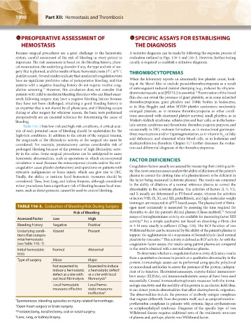
TABLE 116-3. Evaluation of Bleeding Risk During Surgery ogen most commonly is measured by assessing the time required for
15
Risk of Bleeding thrombin to clot the patient’s diluted plasma (Clauss method). Several
assays of transglutaminase activity are available for measuring factor XIII
Assessed Factor Low High
activity, but a simple qualitative test based on dissolving a fibrin clot
16
Bleeding history Negative Positive * in 5 M urea usually is sufficient (Chap. 124). The RCF function of von
Underlying condi- Absent Present Willebrand factor can be measured by the ability of the patient’s plasma to
tions that compro- support the agglutination of a suspension of formaldehyde-fixed normal
mise hemostasis platelets by ristocetin. This activity is defined as RCF activity. As with the
17
(see Table 116–1) coagulation factor assays, the results using patient plasma are compared
Initial hemostatic Normal Abnormal to the results obtained with a normal reference plasma.
tests To determine whether a coagulation factor activity deficiency results
from a quantitative decrease in protein or a qualitative abnormality in the
Type of surgery Minor Major
protein, immunologic assays can be performed using specific polyclonal
Not expected to Expected to induce or monoclonal antibodies to assess the presence of the protein, indepen-
induce a hemostatic a hemostatic defect † dent of its function. Electroimmunoassays, enzyme-linked immunosor-
defect at a site with- at a site with local bent assays (ELISAs), and immunoradiometric assays all have been used
out local fibrinolysis fibrinolysis ‡
successfully. Crossed immunoelectrophoresis measures both the immu-
Local hemostatic Local hemo- nologic reactivity and the mobility of the protein in an electric field; thus,
measures effective static measures it can detect protein abnormalities that affect electrophoretic migration.
ineffective § The abnormalities include the presence of antibody–antigen complexes
that migrate differently from the protein itself, such as antiprothrombin–
* Spontaneous bleeding episodes or injury-related hemorrhage.
† Open heart surgery or brain surgery. prothrombin complexes in patients with systemic lupus erythematosus
or antiphospholipid syndrome. Diagnosis of the specific type of von
‡ Prostatectomy, tonsillectomy, oral or nasal surgery. Willebrand disease requires additional tests of the multimeric structure
§ Liver, lung, or kidney biopsy. of plasma and, perhaps, platelet von Willebrand factor.
Kaushansky_chapter 116_p1985-1992.indd 1990 9/18/15 10:13 AM