
Page 2022 - Williams Hematology ( PDFDrive )
P. 2022
1996 Part XII: Hemostasis and Thrombosis Chapter 117: Thrombocytopenia 1997
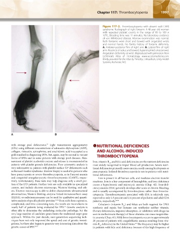
Figure 117–2. Thrombocytopenia with absent radii (TAR)
syndrome. Radiograph of right forearm. A 48-year-old woman
with repeated platelet counts in the range of 85 to 100 ×
9
10 /L. Bleeding time was 11 minutes. No laboratory evidence
of von Willebrand disease. Marrow examination was normal.
Both forearms were short and bowed with angulated wrists
and normal hands. No family history of forearm deformity.
A. Anterior-posterior film of right arm. B. Lateral film of right
arm. Absence of radius and bowed, hypertrophied ulna (arrows).
Angulation deformity at wrist. (Reproduced with permission from
Lichtman’s Atlas of Hematology, www.accessmedicine.com.
Kindly provided for the Atlas by Timothy J. Woodlock, Unity Health
Systems, Rochester, NY.)
A B
with storage pool deficiencies. Light transmission aggregometry NUTRITIONAL DEFICIENCIES
57
(LTA) using different concentrations of adenosine diphosphate (ADP),
collagen, ristocetin, epinephrine, and arachidonic acid is accepted as a AND ALCOHOL-INDUCED
gold standard in diagnosing IPDs, but, again, may be normal in variant THROMBOCYTOPENIA
forms of IPDs and in some patients with storage pool diseases. Mea-
surement of platelet nucleotide content and release is recommended in Iron, vitamin B , and folic acid deficiencies are the nutrient deficiencies
12
patients with platelet granule deficiencies. Flow cytometric analysis is most widely recognized to impair blood cell production. Severe nutri-
very informative in patients with platelet surface GP deficiencies such tional deficiencies primarily cause anemia, rarely causing bicytopenia or
as Bernard-Soulier syndrome. Marrow biopsy is needed in patients who pancytopenia. Isolated thrombocytopenia is rare in patients with nutri-
have pancytopenia or severe thrombocytopenia, as in Fanconi anemia tional deficiencies.
and congenital amegakaryocytic thrombocytopenia (CAMT), respec- Iron is present in all human cells, and mediates electron transfer
tively. Unfortunately, these tests may help diagnose only a small por- reactions. Iron is a key component of hemoglobin, and iron deficiency
tion of the IPD patients. Further tests are only available in specialized causes a hypochromic and microcytic anemia (Chap. 42). Iron-defi-
centers, and include electron microscopy, Western blotting, and oth- ciency anemia (IDA) generally develops after acute or chronic bleeding,
ers. Electron microscopy is able to define characteristic ultrastructural and is usually accompanied by thrombocytosis rather than thrombo-
abnormalities; Western blotting, enzyme-linked immunosorbent assay cytopenia. Thrombocytopenia associated with IDA is relatively rare,
(ELISA), or radioimmunoassay can be used for qualitative and quanti- reported in only 2.3 percent and 2.4 percent of pediatric and adult IDA
tative analysis of specific platelet proteins. 51,56 Even with these expensive, patients, respectively. 58,59
complicated, and time-consuming tests, the results are inconclusive in Cobalamin (vitamin B ) and folate are both required for DNA
12
56
nearly half of patients being evaluated for IPD. Genetic analysis is synthesis and repair, but humans can synthesize neither vitamin.
often able to determine the underlying molecular pathology, but the Dietary deficiencies, impaired absorption, or inhibition with drugs (as
very large number of candidate genes limits the traditional target gene seen in methotrexate therapy) of these vitamins can cause megaloblas-
approach. Within the past decade, next-generation sequencing tech- tic anemia (Chap. 41). Mild thrombocytopenia occurs in approximately
niques have not only improved the speed and cost of genetic investi- 20 percent of patients with megaloblastic anemia resulting from vita-
gations, but have also begun to generate very interesting data about the min B deficiency in the United States. The frequency may be higher
60
12
genetic causes of IPD. 52,53 in patients with folic acid deficiency because of the high frequency of
Kaushansky_chapter 117_p1993-2024.indd 1997 9/21/15 2:31 PM