
Page 2024 - Williams Hematology ( PDFDrive )
P. 2024
1998 Part XII: Hemostasis and Thrombosis Chapter 117: Thrombocytopenia 1999
A B C
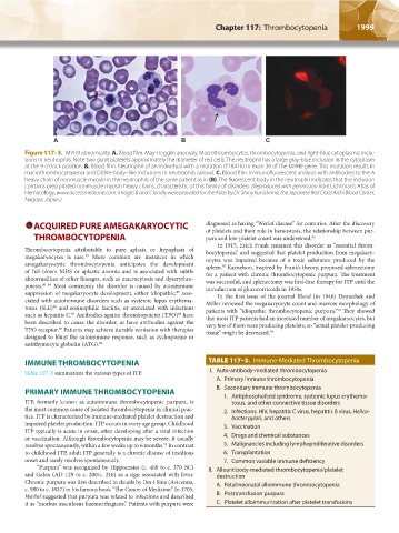
Figure 117–3. MYH9 abnormality. A. Blood film. May-Hegglin anomaly. Macrothrombocytes, thrombocytopenia, and light-blue cytoplasmic inclu-
sions in neutrophils. Note two giant platelets approximately the diameter of red cells. The neutrophil has a large gray-blue inclusion in the cytoplasm
at the 9 o’clock position. B. Blood film. Neutrophil of an individual with a mutation (E1841K) in exon 38 of the MYH9 gene. This mutation results in
macrothrombocytopenia and Döhle-body–like inclusions in neutrophils (arrow). C. Blood film. Immunofluorescent analysis with antibodies to the A
heavy chain of nonmuscle myosin in the neutrophils of the same patient as in (B). The fluorescent body in the neutrophil indicates that the inclusion
contains precipitated nonmuscle myosin heavy chains, characteristic of this family of disorders. (Reproduced with permission from Lichtman’s Atlas of
Hematology, www.accessmedicine.com. Images B and C kindly were provided for the Atlas by Dr. Shinji Kunishima, the Japanese Red Cross Aichi Blood Center,
Nagoya, Japan.)
ACQUIRED PURE AMEGAKARYOCYTIC diagnosed as having “Werlof disease” for centuries. After the discovery
of platelets and their role in hemostasis, the relationship between pur-
THROMBOCYTOPENIA pura and low-platelet count was understood. 92
In 1915, Erich Frank renamed this disorder as “essential throm-
Thrombocytopenia attributable to pure aplasia or hypoplasia of bocytopenia,” and suggested that platelet production from megakary-
megakaryocytes is rare. More common are instances in which ocytes was impaired because of a toxic substance produced by the
79
amegakaryocytic thrombocytopenia anticipates the development spleen. Kaznelson, inspired by Frank’s theory, proposed splenectomy
93
of full-blown MDS or aplastic anemia and is associated with subtle for a patient with chronic thrombocytopenic purpura. The treatment
abnormalities of other lineages, such as macrocytosis and dyserythro- was successful, and splenectomy was first-line therapy for ITP until the
poiesis. 80–84 Most commonly the disorder is caused by autoimmune introduction of glucocorticoids in 1950s.
suppression of megakaryocyte development, either idiopathic, asso- In the first issue of the journal Blood (in 1946) Damashek and
85
ciated with autoimmune disorders such as systemic lupus erythema- Miller reviewed the megakaryocyte count and marrow morphology of
tosus (SLE) and eosinophilic fasciitis, or associated with infections patients with “idiopathic thrombocytopenic purpura.” They showed
86
94
such as hepatitis C. Antibodies against thrombopoietin (TPO) have that most ITP patients had an increased number of megakaryocytes, but
87
88
been described to cause the disorder, as have antibodies against the very few of them were producing platelets, so “actual platelet-producing
TPO receptor. Patients may achieve durable remission with therapies tissue” might be decreased. 94
89
designed to blunt the autoimmune response, such as cyclosporine or
antithymocyte globulin (ATG). 90
IMMUNE THROMBOCYTOPENIA TABLE 117–3. Immune-Mediated Thrombocytopenia
Table 117–3 summarizes the various types of ITP. I. Auto-antibody-mediated thrombocytopenia
A. Primary immune thrombocytopenia
B. Secondary immune thrombocytopenia
PRIMARY IMMUNE THROMBOCYTOPENIA 1. Antiphospholipid syndrome, systemic lupus erythema-
ITP, formerly known as autoimmune thrombocytopenic purpura, is tosus, and other connective tissue disorders
the most common cause of isolated thrombocytopenia in clinical prac- 2. Infections: HIV, hepatitis C virus, hepatitis B virus, Helico-
tice. ITP is characterized by immune-mediated platelet destruction and bacter pylori, and others
impaired platelet production. ITP occurs in every age group. Childhood 3. Vaccination
ITP typically is acute in onset, often developing after a viral infection
or vaccination. Although thrombocytopenia may be severe, it usually 4. Drugs and chemical substances
resolves spontaneously, within a few weeks up to 6 months. In contrast 5. Malignancies including lymphoproliferative disorders
91
to childhood ITP, adult ITP generally is a chronic disease of insidious 6. Transplantation
onset and rarely resolves spontaneously. 7. Common variable immune deficiency
“Purpura” was recognized by Hippocrates (c. 460 to c. 370 BC) II. Alloantibody-mediated thrombocytopenia/platelet
and Galen (AD 129 to c. 200/c. 216) as a sign associated with fever. destruction
Chronic purpura was first described in details by Ibn-i Sina (Avicenna, A. Fetal/neonatal alloimmune thrombocytopenia
c. 980 to c. 1037) in his famous book “The Canon of Medicine.” In 1705,
Werlof suggested that purpura was related to infections and described B. Posttransfusion purpura
it as “morbus maculosus haemorrhagicus.” Patients with purpura were C. Platelet alloimmunization after platelet transfusions
Kaushansky_chapter 117_p1993-2024.indd 1999 9/21/15 2:32 PM