Page 2123 - Williams Hematology ( PDFDrive )
P. 2123

2098           Part XII:  Hemostasis and Thrombosis                                                                                                                                 Chapter 122:  The Vascular Purpuras          2099


                                                                       TABLE 122–1.  Palpable Noninflammatory Purpuric Lesions
                                                                        A.  Dysproteinemias
                                                                           1.  Cryoglobulinemia (see Figs. 122–3 and 122–4)
                                                                           2.  Waldenström hyperglobulinemic purpura (see Fig. 122–5)
                                                                           3.  Light-chain vasculopathy
                                                                           4.  Cryofibrinogenemia
                                                                        B.  Thrombotic
                                                                           1.  Heparin necrosis
                                                                           2.  Warfarin necrosis (see Fig. 122–6)
                                                                           3.  Protein C and protein S deficiencies
                                                                           4.  Paroxysmal nocturnal hemoglobinuria
                                                                           5.  Antiphospholipid syndrome (see Fig. 122–8)
                 A                                                B        6.  Livedoid vasculitis
                                                                        C.  Embolic
                                                                           1.  Cholesterol emboli (see Fig. 122–9)
                                                                           2.  Cutaneous calciphylaxis
                                                                           3.  Emboli from intracardiac thrombi
                                                                        D.  Arthropod bites


                                                                       TABLE 122–2.  Palpable and Nonpalpable Inflammatory
                                                                       Purpuric Lesions
                                                                        A.  Pyoderma gangrenosum (see Fig. 122–7)
                                                                        B.  Sweet syndrome (see Fig. 122–10)
                                                                        C.  Behçet disease
                                                                        D.  Serum sickness (Fig. 122–11)
                                                                        E.  Henoch-Schönlein purpura (see Fig. 122–12)
 A               B                                                      F.  Infections
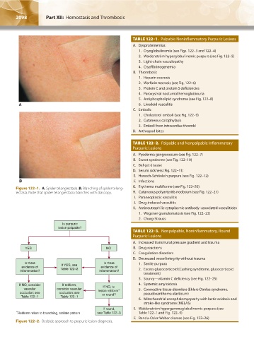
               Figure 122–1.  A. Spider telangiectasia. B. Blanching of spider telang-   G.  Erythema multiforme (see Fig. 122–20)
               iectasia. Note that spider telangiectasia blanches with diascopy.   H.  Cutaneous polyarteritis nodosum (see Fig. 122–21)
                                                                         I.  Paraneoplastic vasculitis
                                                                        J.  Drug-induced vasculitis
                                                                        K.  Antineutrophilic cytoplasmic antibody–associated vasculitides
                                                                           1.  Wegener granulomatosis (see Fig. 122–23)
                                                                           2.  Churg-Strauss
                                     Is purpuric
                                    lesion palpable?
                                                                       TABLE 122–3.  Nonpalpable, Noninflammatory, Round
                                                                       Purpuric Lesions
                                                                        A.  Increased transmural pressure gradient and trauma
                    YES                                   NO            B.  Drug reactions
                                                                        C.  Coagulation disorders
                                                                        D.  Decreased vessel integrity without trauma
                   Is there          If YES, see         Is there          1.  Senile purpura
                  evidence of        Table 122–2        evidence of        2.   Excess glucocorticoid (Cushing syndrome, glucocorticoid
                 inflammation?                         inflammation?
                                                                            treatment)
                                                                           3.  Scurvy—vitamin C deficiency (see Fig. 122–25)
                 If NO, consider     If retiform,        If NO, is         4.  Systemic amyloidosis
                   vascular        consider vascular  lesion retiform ∗      5.   Connective tissue disorders (Ehlers-Danlos syndrome,
                 occlusion; see     occlusion; see      or round?           pseudoxanthoma elasticum)
                  Table 122–1        Table 122–1
                                                                           6.   Mitochondrial encephalomyopathy with lactic acidosis and
                                                                            stroke-like syndrome (MELAS)
                                                         If round,      E.  Waldenström hypergammaglobulinemic purpura (see
                * Retiform refers to branching, stellate pattern  see Table 122–3  Table 122–1 and Fig. 122–5)
                                                                        F.  Rendu-Osler-Weber disease (see Fig. 122–26)
               Figure 122–2.  Bedside approach to purpuric lesion diagnosis.


          Kaushansky_chapter 122_p2097-2112.indd   2098                                                                 9/18/15   10:30 AM
   2118   2119   2120   2121   2122   2123   2124   2125   2126   2127   2128