
Page 2125 - Williams Hematology ( PDFDrive )
P. 2125
2100 Part XII: Hemostasis and Thrombosis Chapter 122: The Vascular Purpuras 2101
Erythematous purpuric lesions associated with homozygous protein C
deficiency can develop within hours of birth and can rapidly progress to
hemorrhagic necrosis. Acquired deficiencies of protein C are associ-
37
ated with autoantibodies to protein C, antibiotics administration, septic
shock, HIV, and liver disease (Chap. 127). Acquired protein S defi-
38
ciency may occur after varicella infection, when it is associated with
39
the generation of antiprotein S immunoglobulins. Protein repletion
with fresh-frozen plasma or protein C concentrate is effective as initial
treatment for protein C deficiency to help clear both cutaneous lesions
and venous occlusion, while lifelong anticoagulant treatment is used to
prevent recurrence. 34,40
Paroxysmal Nocturnal Hemoglobinuria
Paroxysmal nocturnal hemoglobinuria (Chap. 40) is a hematopoietic
clonal disorder resulting in defective production of cell surface-binding
41
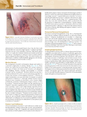
Figure 122–6. Coumadin necrosis. Develops in acral areas and areas proteins. Cutaneous manifestations are secondary to a hypercoag-
of fat deposition such as buttocks or breast. Typically, lesions develop ulable state and include palpable purpura, petechiae, ecchymosis, leg
42
3 to 10 days after initiation of anticoagulant treatment and are caused ulcers, plaques, necrosis, and hemorrhagic bullae. Parvovirus B19 may
by rapid clearing of protein C. The lesions are characterized microscopi- play an etiologic role in the development of cutaneous necrosis. An
43
cally by small-vessel thrombosis. association with pyoderma gangrenosum (Fig. 122–7) and occurrence
44
of purpura fulminans have been described. Histology reveals forma-
45
tion of microvascular fibrin thrombi. 42
administration of unfractionated heparin, but it has also been rarely
described after low-molecular-weight heparin. A delayed-type hyper- Antiphospholipid Syndrome
27
sensitivity reaction to the medication is involved. Skin lesions appear Antiphospholipid syndrome (APS) is a disease characterized by hyper-
within 1 to 2 weeks after treatment initiation and include necrotic pur- coagulability associated with the presence of antibodies against phos-
puric lesions. Development of cutaneous lesions is closely related to pholipids, such as anticardiolipin and lupus anticoagulant (Chap. 131).
46
28
heparin-induced thrombocytopenia (Chap. 118), which involves anti– Approximately 40 percent of patients with APS present with cutane-
platelet factor 4 antibody–mediated platelet aggregation with develop- ous lesions secondary to both large-vessel and microvascular throm-
ment of thrombosis and microvascular occlusion. 27 bosis. Skin manifestations include ecchymosis, livedo reticularis and
47
racemosa, leg ulcerations, bullae, splinter hemorrhages, livedoid vascu-
Warfarin Necrosis lopathy, superficial venous thrombosis, atrophie blanche, and extensive
The development of painful erythematous plaques and nodules is necrosis (Fig. 122–8). 47,48 Presence of livedo reticularis is frequently the
a potential complication of warfarin therapy (Fig. 122–6). These presenting symptom of APS, most commonly when the syndrome is sec-
lesions can rapidly become hemorrhagic and necrotic, leading to ondary to SLE, and its presence commonly precedes vascular events.
49
large areas of infarct with black eschar formation and subsequent Development of acute bullous purpura has been described. Treatment
50
skin sloughing. Purpura, vesicular, maculopapular, or urticarial includes anticoagulant agents with immunosuppressant administra-
eruptions can be encountered. Warfarin-induced necrosis has a tion for associated thrombocytopenia. Prevention of thromboembolic
1
prevalence between 0.01 and 0.1 percent and presents typically 3 to events with aspirin is of uncertain value. 51
10 days after initiation of anticoagulant treatment. 29,30 However, an
atypical presentation can occur much later, for example, in a patient
with protein S deficiency. 31,32 Although warfarin necrosis tends to
develop in areas of greatest fat deposition, such as breasts, thighs,
and buttocks, acral areas, including penis, fingers, and toes, can also
be involved. Warfarin necrosis results from the rapid decrease of
33
vitamin K–dependent coagulation factors of relatively short half-life,
such as proteins C and S, while longer-lasting coagulation factors,
such as factor II and factor X, are not yet decreased, resulting in a
net procoagulant state. Microvascular occlusion of small dermal and
subcutaneous vessels by fibrin deposits is seen on histologic analysis,
but true vasculitis is infrequent. Treatment involves prompt cessation
29
of the vitamin K antagonist, along with administration of heparin and
vitamin K, and occasionally surgical debridement. Because patients
with protein C or S deficiency are at increased susceptibility to warfarin
necrosis, heparin should always be administered in these patients prior
to initiation of Coumadin. 34
Proteins C and S Deficiencies Figure 122–7. Pyoderma gangrenosum. A large number of systemic
Clinical manifestations of proteins C and S deficiencies include venous diseases are associated with pyoderma gangrenosum, including inflam-
matory bowel diseases, hematologic and solid malignancies, and rheu-
thromboembolism, warfarin-induced skin necrosis, and neonatal pur- matologic disorders. Microscopically, the lesions are characterized by
pura fulminans (Chap. 129). Congenital and acquired deficiencies in central necrotizing, neutrophilic infiltration, and a surrounding perivas-
these proteins can lead to palpable necrotic purpura and ecchymosis. 35,36 cular and intramural lymphocytic infiltration.
Kaushansky_chapter 122_p2097-2112.indd 2100 9/18/15 10:30 AM