
Page 2127 - Williams Hematology ( PDFDrive )
P. 2127
2102 Part XII: Hemostasis and Thrombosis Chapter 122: The Vascular Purpuras 2103
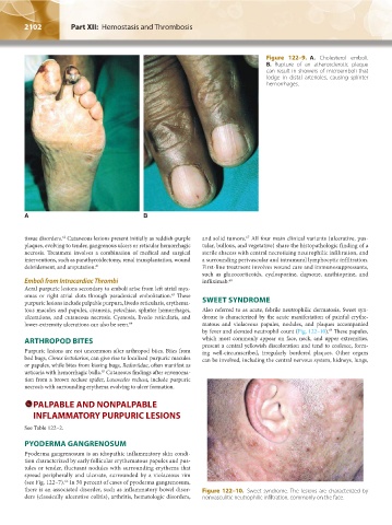
Figure 122–9. A. Cholesterol emboli.
B. Rupture of an atherosclerotic plaque
can result in showers of microemboli that
lodge in distal arterioles, causing splinter
hemorrhages.
A B
tissue disorders. Cutaneous lesions present initially as reddish-purple and solid tumors. All four main clinical variants (ulcerative, pus-
67
62
plaques, evolving to tender, gangrenous ulcers or reticular hemorrhagic tular, bullous, and vegetative) share the histopathologic finding of a
necrosis. Treatment involves a combination of medical and surgical sterile abscess with central necrotizing neutrophilic infiltration, and
interventions, such as parathyroidectomy, renal transplantation, wound a surrounding perivascular and intramural lymphocytic infiltration.
debridement, and amputation. 61 First-line treatment involves wound care and immunosuppressants,
such as glucocorticoids, cyclosporine, dapsone, azathioprine, and
Emboli from Intracardiac Thrombi infliximab. 68
Acral purpuric lesions secondary to emboli arise from left atrial myx-
omas or right atrial clots through paradoxical embolization. These
63
purpuric lesions include palpable purpura, livedo reticularis, erythema- SWEET SYNDROME
tous macules and papules, cyanosis, petechiae, splinter hemorrhages, Also referred to as acute, febrile neutrophilic dermatosis, Sweet syn-
ulcerations, and cutaneous necrosis. Cyanosis, livedo reticularis, and drome is characterized by the acute manifestation of painful erythe-
lower-extremity ulcerations can also be seen. 64 matous and violaceous papules, nodules, and plaques accompanied
69
by fever and elevated neutrophil count (Fig. 122–10). These papules,
ARTHROPOD BITES which most commonly appear on face, neck, and upper extremities,
present a central yellowish discoloration and tend to coalesce, form-
Purpuric lesions are not uncommon after arthropod bites. Bites from ing well-circumscribed, irregularly bordered plaques. Other organs
bed bugs, Cimex lectularius, can give rise to localized purpuric macules can be involved, including the central nervous system, kidneys, lungs,
or papules, while bites from kissing bugs, Reduviidae, often manifest as
65
urticaria with hemorrhagic bulla. Cutaneous findings after envenoma-
tion from a brown recluse spider, Loxosceles reclusa, include purpuric
necrosis with surrounding erythema evolving to ulcer formation.
PALPABLE AND NONPALPABLE
INFLAMMATORY PURPURIC LESIONS
See Table 122–2.
PYODERMA GANGRENOSUM
Pyoderma gangrenosum is an idiopathic inflammatory skin condi-
tion characterized by early follicular erythematous papules and pus-
tules or tender, fluctuant nodules with surrounding erythema that
spread peripherally and ulcerate, surrounded by a violaceous rim
(see Fig. 122–7). In 50 percent of cases of pyoderma gangrenosum,
66
there is an associated disorder, such as inflammatory bowel disor- Figure 122–10. Sweet syndrome. The lesions are characterized by
ders (classically ulcerative colitis), arthritis, hematologic disorders, nonvasculitic neutrophilic infiltration, commonly on the face.
Kaushansky_chapter 122_p2097-2112.indd 2102 9/18/15 10:30 AM