
Page 2124 - Williams Hematology ( PDFDrive )
P. 2124
2098 Part XII: Hemostasis and Thrombosis Chapter 122: The Vascular Purpuras 2099
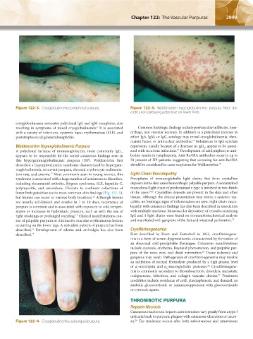
Figure 122–3. Cryoglobulinemia: peripheral purpura. Figure 122–5. Waldenström hyperglobulinemic purpura. Note dis-
crete and coalescing petechiae on lower limb.
cryoglobulinemia associates polyclonal IgG and IgM complexes, also
resulting in symptoms of mixed cryoglobulinemia. It is associated Common histologic findings include perivascular infiltrates, hem-
6
with a variety of infections, systemic lupus erythematous (SLE), and orrhage, and vascular necrosis. In addition to a polyclonal increase in
poststreptococcal glomerulonephritis. either IgA, IgM, or IgG, serology may reveal cryoglobulinemia, rheu-
matoid factor, or antinuclear antibodies. Imbalances in IgG subclass
18
Waldenström Hyperglobulinemic Purpura expression, usually because of a decrease in IgG , appear to be associ-
2
A polyclonal increase of immunoglobulins, most commonly IgG , ated with recurrent infections. Development of antilymphocyte anti-
17
1
appears to be responsible for the varied cutaneous findings seen in bodies results in lymphopenia. Anti-Ro/SSA antibodies occur in up to
this hypergammaglobulinemic purpura (HP). Waldenström first 78 percent of HP patients, suggesting that screening for anti-Ro/SSA
described a hyperproteinemic syndrome characterized by hypergam- should be considered in cases suspicious for Waldenström. 19
maglobulinemia, recurrent purpura, elevated erythrocyte sedimenta-
tion rate, and anemia. Most commonly seen in young women, this Light-Chain Vasculopathy
13
syndrome is associated with a large number of autoimmune disorders, Precipitates of immunoglobulin light chains that form crystalline
including rheumatoid arthritis, Sjögren syndrome, SLE, hepatitis C, deposits in the skin cause hemorrhagic palpable purpura. A nonamyloid
polymyositis, and sarcoidosis. Discrete to confluent collections of monoclonal light chain of predominant κ type is involved in two-thirds
lower limb petechiae are its most common skin findings (Fig. 122–5), of the cases. 20,21 Crystalline deposits are present in the skin and other
but lesions can occur in various body locations. Although lesions tissues. Although the clinical presentation may mimic a systemic vas-
14
are usually self-limited and resolve in 7 to 10 days, recurrence of culitis, no histologic signs of inflammation are seen. Light-chain vascu-
purpura is common and is associated with exposure to cold temper- lopathy with cutaneous findings has also been described in association
atures or increases in hydrostatic pressure, such as with the use of with multiple myeloma. Intravascular deposition of crystals containing
tight stockings or prolonged standing. Clinical manifestations con- IgG and λ light chains were found on immunohistochemical analysis
15
sist of palpable purpura or diminutive macular erythematous lesions and manifested with gangrene of the feet and intestinal perforation. 22
occurring on the lower legs. A reticulate pattern of purpura has been
described. Development of edema and arthralgia has also been Cryofibrinogenemia
16
described. 17 First described by Korst and Kratochvil in 1955, cryofibrinogene-
mia is a form of serum dysproteinemia characterized by formation of
an abnormal cold-precipitable fibrinogen. Cutaneous manifestations
include cyanosis, erythema, Raynaud phenomenon, and palpable pur-
pura of the nose, ears, and distal extremities. Tissue ischemia and
23
gangrene may result. Pathogenesis of cryofibrinogenemia may involve
an inhibition of normal fibrinolysis produced by a high plasma level
of α -antitripsin and α -macroglobulin proteases. Cryofibrinogene-
24
1
2
mia is commonly secondary to thromboembolic disorders, metastatic
malignancies, infections, and collagen vascular disease. Treatment
25
modalities include avoidance of cold, plasmapheresis, and danazol, an
anabolic glucocorticoid, or immunosuppression with glucocorticoids
or cytotoxic agents.
THROMBOTIC PURPURA
Heparin Necrosis
Cutaneous reactions to heparin administration vary greatly from a type I
urticarial rash to purpuric plaques with cutaneous ulceration or necro-
26
Figure 122–4. Cryoglobulinemia: subungual purpura. sis. The syndrome occurs after both subcutaneous and intravenous
Kaushansky_chapter 122_p2097-2112.indd 2099 9/18/15 10:30 AM