
Page 2126 - Williams Hematology ( PDFDrive )
P. 2126
2100 Part XII: Hemostasis and Thrombosis Chapter 122: The Vascular Purpuras 2101
A B
C
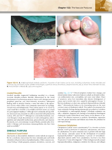
Figure 122–8. A. Antiphospholipid antibody syndrome. A number of skin lesions can be seen, including ecchymosis, livedo reticularis and
racemosa, leg ulcerations, bullae, splinter hemorrhages, superficial venous thrombosis, atrophie blanche, and, as shown here, extensive necrosis.
B. Anticardiolipin antibody. C. Lupus anticoagulant.
56
Livedoid Vasculitis nodules (Fig. 122–9). Clinical symptoms include fever, myalgia, and
Livedoid vasculitis (segmental hyalinizing vasculitis) is a chronic altered mental status. Laboratory features include an elevated erythro-
recurrent thrombo-occlusive disorder characterized by the initial cyte sedimentation rate, eosinophilia, and acute renal failure. Onset
development of erythematous purpuric lesions with telangiectasis and of symptoms varies from immediate after physical dislodgement of
1
peripheral petechiae, and lower-extremity ulcerations. Subsequent plaque, up to months later when caused by anticoagulant therapy. A
healing leads to atrophie blanche, a term that refers to the appear- blue toe syndrome is, in fact, rare, and most atheroemboli are clinically
57
ance of ivory-white stellate scars commonly surrounded by hyperpig- silent. Atherosclerotic lesions in the descending aorta are the most
mented areas and telangiectasia. These lesions appear to be caused by common source of cholesterol emboli. This explains the propensity for
small-vessel fibrin thrombi in the middle and lower dermis as a result lower-extremity findings during intravascular procedures or initiation
56
of a procoagulant tendency. Although most commonly arising with- of thrombolytic or anticoagulant therapy. Histologic evaluation can
52
out associated cause, livedoid vasculitis is associated with polyarteritis offer a definitive diagnosis with findings of intraluminal birefringent
nodosa, APS, and SLE. 53,54 Although not consistently beneficial, com- cholesterol crystals within blood vessel lumen, in the absence of vas-
58
mon therapies include discontinuation of oral contraceptives, antico- culitis. No effective treatment is available. Nevertheless, supportive
agulation and antiplatelet medications, glucocorticoids, and dapsone. care with proper hydration and dialysis may lessen the potential for
Ketanserin, an S serotoninergic receptor blocker, psoralen plus ultra- end-organ damage.
2
violet A therapy, and intravenous immunoglobulins also have been
used successfully. 55 Cutaneous Calciphylaxis
Calciphylaxis (calcific uremic arteriolopathy) is a thrombo-occlusive
59
EMBOLIC PURPURA disorder involving formation of cutaneous, subcutaneous, and vascu-
lar calcifications. It is most commonly seen in patients with end-stage
Cholesterol Crystal Emboli renal disease, classically caused by the development of secondary
Also known as atheroemboli, cholesterol crystal emboli are responsi- hyperparathyroidism. Approximately 4 percent of hemodialysis-
60
ble for a syndrome characterized by lower extremity pain and livedo dependent patients suffer from calciphylaxis. Survival is less than
reticularis with preservation of peripheral pulses. Other common cuta- 50 percent at 5 years after diagnosis. Other etiologies include primary
61
neous findings include gangrene, purpura, ulcerations, cyanosis, and hyperparathyroidism, malignancy, alcoholic liver disease, and collagen
Kaushansky_chapter 122_p2097-2112.indd 2101 9/18/15 10:30 AM