
Page 2128 - Williams Hematology ( PDFDrive )
P. 2128
2102 Part XII: Hemostasis and Thrombosis Chapter 122: The Vascular Purpuras 2103
and bones. Classically more prominent in middle-aged women, this HENOCH-SCHÖNLEIN PURPURA
70
syndrome associates a complex cytokine dysregulation. Other mani- Henoch-Schönlein purpura (HSP), is a predominantly pediatric vas-
festations include respiratory and urinary infections and autoimmune culitic syndrome characterized by the acute onset of abdominal pain
disorders (including rheumatoid arthritis), SLE, inflammatory bowel and lower-extremity eruption of diffuse urticarial plaques and palpa-
disease). Histologic analysis shows a distinct nonvasculitic neutrophilic ble purpura. It was first described in 1801 by Dr. William Heberden.
78
infiltrate in the superficial dermis with dermal edema. Systemic glu- HSP predominantly affects patients 2 to 20 years of age, 90 percent
cocorticoid treatment is the standard treatment, while clofazimine, of patients being younger than 10 years old. Several environmental
79
dapsone, colchicine, indomethacin, and cyclosporine have also been triggers precede HSP onset, such as viral (upper respiratory infec-
used successfully. 71
tions, hepatitis B virus, HCV, parvovirus B19, and HIV) and bacterial
(Streptococcus spp., Staphylococcus aureus, and Salmonella spp.) infec-
BEHÇET DISEASE tions in children. Adult disease may be precipitated by medications
Besides its classification as a neutrophilic dermatosis, Behçet disease (nonsteroidal antiinflammatory drugs [NSAIDs], angiotensin-convert-
is also an inflammatory disorder that affects multiple organ systems. ing enzyme inhibitors, and antibiotics), food allergies, vaccinations,
80
Clinical features include chronic and relapsing cutaneous manifesta- and insect bites. The pathogenesis of HSP leukocytoclastic vasculitis
tions, such as palpable purpura, infiltrative erythema, and papulopus- is complex. It appears to involve IgA immune complex and comple-
1
tular lesions, as well as oral mucosal and genital ulcers, arthralgias, and ment deposition on vessel walls. Elevated values of thrombomodulin,
gastrointestinal and central nervous system involvement. Genetic tissue plasminogen activator, and plasminogen activator inhibitor-1
72
studies show an association between Behçet disease and human leu- appear to correlate with endothelial injury and fibrinolytic activity in
kocyte antigen B51. Histologic features include leukocytoclastic or the acute phase of HSP. 81
73
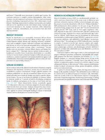
lymphocytic vasculitis, hence its previous classification as a vasculitis. Cutaneous eruptions often begin acutely as urticarial papules and
Antitumor necrosis factor-α directed therapies (infliximab, etanercept), plaques evolving to petechiae, ecchymoses, and palpable and nonpal-
interferon-α, immunosuppressive and immunomodulatory agents such pable purpura over the lower extremities and buttocks (Fig. 122–12).
as thalidomide, intravenous immunoglobulin, and even stem cell trans- Palpable purpura is a universal finding, being present in one series
82
plantation are used in Behçet disease. 74,75 in 98.6 percent of patients. Clinically, lesions may take the form of
retiform or patterned purpura, presence of a retiform edge of various
83
SERUM SICKNESS inflammatory lesions, or skin necrosis. Other common manifestations
include localized subcutaneous edema, glomerulonephritis, arthritis,
Serum sickness reflects the clinical manifestations of immune complex and (severe) abdominal pain.
formation and deposition. Cutaneous lesions such as urticarial and In spite of its chronic relapsing pattern, the long-term evolution is
morbilliform eruptions predominate, though palpable purpura and benign in the majority of patients. The self-limited course of HSP may
82
erythema multiforme can also be encountered. Serum sickness asso- be contributed by an enhanced apoptosis of immune cells, which dimi-
ciated with infection or medical therapy can result in specific charac- nishes the severity of the acute inflammatory response. Consequently,
84
teristic lesions. The use of antithymocyte globulin for marrow failure, treatment is frequently supportive. Immunosuppressive drugs, including
for instance, results in 75 percent of patients developing serpiginous glucocorticoids, are typically reserved for cases with renal involvement.
78
bands of erythema and purpura on the sides of their hands and feet
(Fig. 122–11). These characteristic lesions consistently appear
76
1 to 2 days prior to the onset of systemic symptoms of serum sickness,
which include fever and malaise. Analysis of biopsies by direct immu-
nofluorescence reveals deposition of IgM, IgE, IgA, and C3. This depo-
sition appears to activate neutrophils leading to release of lysosomal
enzymes and the development of dermal vasculitis. 77
Figure 122–12. Henoch-Schönlein purpura. Urticarial papules and
Figure 122–11. Serum sickness caused by antithymocyte globulin. plaques can evolve into palpable purpura. The lesions are characterized
The lesions consist of immunoglobulins and neutrophils. by leukocytoclastic vasculitis.
Kaushansky_chapter 122_p2097-2112.indd 2103 9/18/15 10:30 AM