
Page 306 - Williams Hematology ( PDFDrive )
P. 306
280 Part IV: Molecular and Cellular Hematology Chapter 19: The Inflammatory Response 281
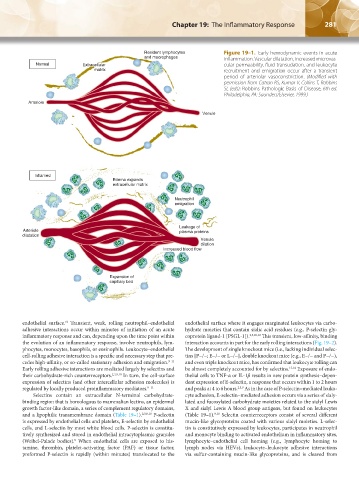
Resident lymphocytes Figure 19–1. Early hemodynamic events in acute
and macrophages inflammation. Vascular dilatation, increased microvas-
Normal Extracellular cular permeability, fluid transudation, and leukocyte
matrix recruitment and emigration occur after a transient
period of arteriolar vasoconstriction. (Modified with
permission from Cotran RS, Kumar V, Collins T, Robbins
SL (eds): Robbins Pathologic Basis of Disease, 6th ed.
Philadelphia, PA: Saunders/Elsevier, 1999.)
Arteriole
Venule
Inflamed
Edema expands
extracellular matrix
Neutrophil
emigration
Leakage of
Arteriole plasma proteins
dilatation
Venule
dilation
Increased blood flow
Expansion of
capillary bed
endothelial surface. Transient, weak, rolling neutrophil–endothelial endothelial surface where it engages marginated leukocytes via carbo-
10
adhesive interactions occur within minutes of initiation of an acute hydrate moieties that contain sialic acid residues (e.g., P-selectin gly-
inflammatory response and can, depending upon the time point within coprotein ligand-1 [PSGL-1]). 2,3,10,12 This transient, low-affinity, binding
the evolution of an inflammatory response, involve neutrophils, lym- interaction accounts in part for the early rolling interactions (Fig. 19–2).
phocytes, monocytes, basophils, or eosinophils. Leukocyte–endothelial The development of single knockout mice (i.e., lacking individual selec-
cell-rolling adhesive interaction is a specific and necessary step that pre- tins [P−/−; E−/− or L−/−], double knockout mice (e.g., E−/− and P−/−),
cedes high-affinity, or so-called stationary adhesion and emigration. 9–11 and even triple knockout mice, has confirmed that leukocyte rolling can
Early rolling adhesive interactions are mediated largely by selectins and be almost completely accounted for by selectins. 13,14 Exposure of endo-
their carbohydrate-rich counterreceptors. 2,3,9,10 In turn, the cell-surface thelial cells to TNF-α or IL-1β results in new protein synthesis–depen-
expression of selectins (and other intercellular adhesion molecules) is dent expression of E-selectin, a response that occurs within 1 to 2 hours
regulated by locally produced proinflammatory mediators. 9–11 and peaks at 4 to 6 hours. 2,3,9 As in the case of P-selectin–mediated leuko-
Selectins contain an extracellular N-terminal carbohydrate- cyte adhesion, E-selectin–mediated adhesion occurs via a series of sialy-
binding region that is homologous to mammalian lectins, an epidermal lated and fucosylated carbohydrate moieties related to the sialyl Lewis
growth factor-like domain, a series of complement regulatory domains, X and sialyl Lewis A blood group antigens, but found on leukocytes
and a lipophilic transmembrane domain (Table 19–1). 2,3,9,10 P-selectin (Table 19–1). Selectin counterreceptors consist of several different
9,10
is expressed by endothelial cells and platelets, E-selectin by endothelial mucin-like glycoproteins coated with various sialyl moieties. L-selec-
cells, and L-selectin by most white blood cells. P-selectin is constitu- tin is constitutively expressed by leukocytes, participates in neutrophil
tively synthesized and stored in endothelial intracytoplasmic granules and monocyte binding to activated endothelium in inflammatory sites,
(Weibel-Palade bodies). When endothelial cells are exposed to his- lymphocyte–endothelial cell homing (e.g., lymphocyte homing to
9
tamine, thrombin, platelet-activating factor (PAF) or tissue factor, lymph nodes via HEVs), leukocyte–leukocyte adhesive interactions
preformed P-selectin is rapidly (within minutes) translocated to the via sulfur-containing mucin-like glycoproteins, and is cleaved from
Kaushansky_chapter 19_p0279-0292.indd 281 9/17/15 5:51 PM