
Page 309 - Williams Hematology ( PDFDrive )
P. 309
284 Part IV: Molecular and Cellular Hematology Chapter 19: The Inflammatory Response 285
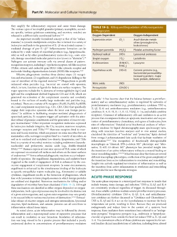
they amplify the inflammatory response and cause tissue damage. TABLE 19–2. Killing and Degradation of Microorganisms
The various types of neutrophil granules (primary azurophilic, second-
ary specific, tertiary gelatinase-containing, and secretory vesicles) are in Phagocytes
released in a differentially coordinated fashion. 21,22 Oxygen-Dependent Oxygen-Independent
An important recently formulated concept is that of the “inflam- Superoxide anion (O ) Arachidonate metab-
−
masome,” a cytosolic multiprotein complex that is formed in a variety of 2 olites (prostaglandins,
leukocytes and leads to the generation of IL-1β via activated caspase-1– leukotrienes)
23
mediated cleavage of pro–IL-1β. Inflammasome formation can be Hydrogen peroxide (H O ) Platelet-activating factor
induced by a wide variety of microbial products (e.g., lipopolysaccha- 2 2
ride) as well as by other proinflammatory molecules (e.g., urate crys- Hydroxyl radical (HO•) Lysosomal proteases
23
tals, damage-associated molecular patterns also known as “alarmins”). Singlet oxygen ( O ) Lactoferrin
1
2
Pathogens can activate immune cells via several classes of pattern- N-chloramines (R-NHC1, Lysozyme
recognition receptors, including C-type lectin receptors, toll-like receptors R-NCl )
(TLRs), retinoic acid–inducible gene (RIG) 1-like cytosolic receptor and 2
nucleotide-binding oligomerization domain (NOD)-like receptors. 23,24 Hypohalous acids (HO-X) Cationic proteins (e.g.,
Effective phagocytosis involves three distinct steps: (1) recogni- bactericidal permeability-
increasing protein, major
tion and attachment, (2) engulfment, and (3) degradation (killing in the basic protein, defensins)
case of microbes) of the ingested material. 21,22 Phagocytosis is greatly
enhanced when particles (e.g., bacteria) are coated with opsonins, Nitric oxide (NO)
which, in turn, function as ligands for leukocyte surface receptors. The Peroxynitrite (ONOO )
−
major opsonins include the Fc domains of immunoglobulin (Ig) G and
IgM and the complement-derived fragments C3b and iC3b, which are
generated via activation of the complement cascades and covalently and cells. It has become clear that the balance between a proinflam-
5
bound to the surfaces of nearby large molecules and particles (e.g., matory and an antiinflammatory milieu is regulated by networks of
microbes). There are a variety of Fc receptors (FcγRI, FcγRII, FcγRIIIB, proinflammatory mediators (e.g., proinflammatory cytokines: TNF-α,
etc.) and complement receptors (e.g., CR1, CR3, CR4) that specifically IL-1β, IL-6) and antiinflammatory mediators (e.g., antiinflammatory
engage their respective opsonins when the latter coat foreign partic- cytokines: IL-4, IL-10, IL-11, IL-13, TGFβ, IL-1ra, and soluble cytokine
ulates. In addition to facilitating receptor-mediated phagocytosis of receptors). Clearance of inflammatory cells and mediators is an active
6
opsonized particles, Fc receptors trigger cell activation with the atten- process that encompasses leukocyte apoptosis, inactivation and seques-
dant release of granular constituents and the generation of reactive oxy- tration of proinflammatory chemokines, and egress of leukocytes from
6
gen intermediates. Other important recognition molecules expressed sites of inflammation. Detailed serial biochemical analyses of inflam-
5
by leukocytes include integrins, the C1q receptor, mannose receptors, matory exudates by liquid chromatography-mass spectroscopy have,
scavenger receptors and TLRs. 6,24,25 Mannose receptors bind to man- along with structure–function analyses and in vivo animal studies,
nose and fucose moieties, which are present on some microbes but not elucidated the identities of “resolvins” and “protectins,” lipids derived
mammalian cells; scavenger receptors bind to a variety of microbes, as from omega-3 polyunsaturated fatty acids that facilitate resolution of
well as to oxidized and acetylated low-density lipoproteins; and TLRs inflammation. 5,26,27 The recognition of functionally distinguishable
bind to a variety of microbial moieties including endotoxins (lipopoly- macrophages as “classical, IFN-γ–driven M1” phenotype and “alter-
saccharide) and prokaryotic nucleic acids (e.g., double-stranded native, IL-4/IL-13–driven M2” phenotype has provided insight into
RNA). 6,24,25 Humans express at least nine species of TLR, some of which the transition of an active inflammatory milieu to a wound-healing or
are expressed on external cell surfaces and others on the inner surfaces tissue-remodeling milieu. 5,28,2928 It has become clear that there are several
of endosomes. 24,25 Some enhanced phagocytic reactions occur indepen- different macrophage phenotypes, a reflection of the great complexity of
dently of opsonins. The engulfment, degranulation, and oxidative burst the transition from active inflammation to resolution and remodeling.
triggered as the result of engagement of FcR is enhanced by the con- Insight into actively regulated termination of inflammation as well as
current engagement of complement receptors. In some circumstances, the transition from active inflammation to resolution and remodeling
engulfment is enhanced by the simultaneous binding of the leukocyte has provided for new therapeutic strategies.
to specific extracellular matrix molecules (e.g., fibronectin) or soluble
cytokines. Engulfment results in the formation of phagosomes, which
fuse with lysosomes to form phagolysosomes in which foreign particles ACUTE-PHASE RESPONSE
are oxidized and degraded. Numerous mechanisms for killing and/or The acute-phase response is a stereotyped host response to insults that
degradation of microbes have been elucidated (Table 19–2). Although include trauma, tissue damage, and infection. TNF-α, IL-1β, and IL-6
3
these mechanisms are classified as either oxygen-dependent or oxygen- are consistently produced regardless of trigger. As discussed through-
independent, both types of processes may be involved in the destruction out, these soluble cytokines mediate several proinflammatory processes.
of a given microorganism, and a given microorganism may vary greatly (Proinflammatory cytokines TNF-α, IL-1β, IL-6, and antiinflamma-
in its susceptibility to various mechanisms of destruction. 6,21,22 Extracel- tory cytokines are further discussed in “Cytokines and Chemokines”.)
lular release of reactive oxygen and nitrogen intermediates, lysosomal TNF-α, IL-1β and IL-6 act on the hypothalamus to increase the body
enzymes, lipid mediators, and cationic proteins can all contribute to temperature set point, resulting in fever. Because they are produced
inflammation-related tissue injury. endogenously and induce fever in the context of a systemic host
As noted above, acute inflammation may be followed by chronic response, these mediators have sometimes been referred to as “endoge-
inflammation and a superimposed series of reparative processes that nous pyrogens.” Exogenous pyrogens (e.g., endotoxin or lipopolysac-
can result in resolution or scar formation. Resolution of inflamma- charide) originate from outside the host but induce TNF-α, IL-1β, and
tion was long viewed to be a passive process that included a poorly IL-6. The downstream effects of these cytokines are responsible for sev-
understood decline in concentrations of proinflammatory mediators eral familiar clinical manifestations of infection, including fever, altered
Kaushansky_chapter 19_p0279-0292.indd 284 9/17/15 5:51 PM