
Page 311 - Williams Hematology ( PDFDrive )
P. 311
286 Part IV: Molecular and Cellular Hematology Chapter 19: The Inflammatory Response 287
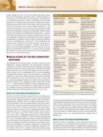
marked changes not only in the local cytokine–chemokine milieu, TABLE 19–3. Inflammatory Mediator Systems
but also in the lipid milieu. Specifically, there is a shift from high local
concentrations of proinflammatory prostaglandins and leukotrienes Mediator System Source Major Actions
to antiinflammatory lipoxins, resolvins, and protectins. Basic lipoxin Reactive oxygen Leukocytes, Tissue damage through
biochemistry is outlined later in the section “Inflammatory Lipids”. intermediates (O , endothelial cells cytolysis, matrix deg-
−
2
Resolvins and protectins, derived from omega-3 polyunsaturated fatty H O , HOX, HO) radation, activation of
2
2
acids, each encompass several classes of related molecules. 5,26,27 The complement, and gen-
synthesis of both resolvins and protectins occurs through enzymatic eration of chemotactic
pathways; the mediators themselves exert their effects through specific lipids
receptors on neutrophils, macrophages and dendritic cells. 5,26,27 Antiin- Reactive nitrogen Monocytes, Cytostasis of cells, inhi-
flammatory mechanisms exerted by resolvins and protectins are diverse intermediates (NO, macrophages, bition of DNA synthesis,
−
−
−
and include enhancement of macrophage-mediated clearance of apop- ONOO , NO , NO ) lymphocytes, inhibition of mitochon-
2
3
totic neutrophils, upregulation of cell-surface CCR5 which sequesters endothelial cells drial respiration, and
proinflammatory chemokines, and the shift of biochemical pathways formation of OH
toward an antiinflammatory phenotype. 3,26,27 The identification and Lysosomal granule Neutrophils, Tissue damage through
characterization of both resolvins and protectins has laid the ground- constituents (pro- monocytes proteolysis, matrix deg-
work for new therapeutic strategies to modulate inflammation. teases, lysozyme, radation, and catalysis
5,26
Efficacy of several “proresolving lipids” has been reported in animal lactoferrin, cationic of oxidant-generating
models of inflammation. Resolvin E1, a synthetic resolvin analogue proteins) reactions
5
(RX-100045), and LXA4-based compounds are under investigation in Cytokines and Monocytes, mac- Cell activation, induc-
several human inflammatory diseases. 5 chemokines (TNF, rophages, and tion of adhesion,
IL-1, IL-8, MCP-1, endothelial cells chemotaxis, fever, and
REGULATORS OF THE INFLAMMATORY etc.) Leukocytes, acute-phase response
Platelet-activating
Vascular permeability
RESPONSE factor endothelial cells and cell activation
The foregoing sections have provided a conceptual framework for the Arachidonic acid Cell membranes Coagulation, vasodila-
inflammatory response, specifically, the hemodynamic alterations, metabolites (pros- (endothelial tion, vascular perme-
taglandins, 5-HPETE, cells, platelets,
ability, cell activation,
mechanisms of specific leukocyte–endothelial adhesive interactions, leukotrienes) leukocytes) and chemotaxis
chemotaxis, leukocyte activation, phagocytosis, intracellular microbial
killing mechanisms, active termination/resolution of the acute inflam- Kinins (bradykinin, Plasma Pain, vascular permea-
matory response, and the contributions of M1 and M2 macrophages kallikrein) bility, and vasodilation
to inflammation and tissue repair. The many steps that constitute this Vasoactive amines Platelets, mast Vascular permeability,
paradigm are regulated by soluble mediators produced by endothelial (serotonin, cells, and induction of adhesion
cells and leukocytes at a site of inflammation, by other resident cells histamine) basophils
(e.g., tissue macrophages, fibroblasts, mast cells) and as byproducts of Complement Plasma, Chemotaxis, vascular
bloodborne proteins (e.g., complement system, coagulation cascade; macrophages permeability, and cell
Table 19–3). There are many examples of “crosstalk” among regulatory sys- activation
tems (e.g., proteinase-activated receptors), complex regulatory networks Coagulation Plasma Chemotaxis, vascular
(e.g., proinflammatory and antiinflammatory cytokine balance), and plei- permeability, and com-
otropism exhibited by individual mediators (e.g., TNF-α and IL-1β). plement activation
REACTIVE OXYGEN INTERMEDIATES 5-HPETE, 5 hydroperoxyeicosatetraenoic acid; IL, interleukin; MCP-1,
monocyte chemoattractant protein-1; TNF, tumor necrosis factor.
Since the early 1970s it has been recognized that activated phagocytes
exhibit a transient but marked increase in oxygen consumption and the
mechanistically coupled generation of reduced oxygen metabolites. many types of parenchymal cells. Implicated biochemical mechanisms
32
33
Although small quantities of reactive oxygen intermediates are pro- include lipid peroxidation, formation of carbonyl moieties and nitrosy-
duced as byproducts of several metabolic pathways, the chief source is lation products, intracellular enzyme inactivation, protein oxidation,
the leukocyte cytosol and membrane-associated NADPH oxidase, an and oxidant-mediated DNA damage. Reactive oxygen intermediates
enzyme complex that is defective in most patients with chronic gran- (e.g., O ) can also undergo reactions with reactive nitrogen intermedi-
−
2
ulomatous disease (Chap. 66). Reactive oxygen intermediates include ates (e.g., NO; see “Reactive Nitrogen Intermediates” below) to generate
superoxide anion (O ), hydrogen peroxide (H O ), hydroxyl radical toxic NO derivatives. Within limits, host cells are protected by anti-
−
33
2
2
2
(HO•), and singlet oxygen ( O ). These reduced oxygen products oxidant defense systems (e.g., superoxide dismutase, catalase, reduced
32
1
2
play a major role in intraphagolysosomal killing of microorganisms, glutathione). 33
and when released extracellularly, are directly or indirectly responsible
for a variety of proinflammatory processes, including endothelial cell
lysis, extracellular matrix degradation, activation of latent proteolytic REACTIVE NITROGEN INTERMEDIATES
enzymes (collagenase, gelatinase), inactivation of antiproteases, inter- Described in 1980 as “endothelium-derived relaxing factor,” NO is the
action with metabolites of l-arginine, and generation of chemotactic soluble, gaseous, short-acting biosynthetic product of l-arginine, O ,
2
34
factors from arachidonic acid and the complement component, C5. NADPH, and NO synthase (NOS). As suggested by its original name,
33
In addition to their role in endothelial cytotoxicity, reactive oxygen NO mediates vascular smooth muscle relaxation. NO binds to the heme
intermediates are cytotoxic to fibroblasts, erythrocytes, tumor cells and moiety of guanylyl cyclase to trigger the generation of intracytoplasmic
Kaushansky_chapter 19_p0279-0292.indd 286 9/17/15 5:51 PM