
Page 313 - Williams Hematology ( PDFDrive )
P. 313
288 Part IV: Molecular and Cellular Hematology Chapter 19: The Inflammatory Response 289
trauma) can induce macrophages (and other cell types) to secrete TNF- of chemokines is based on the locations of N-terminal cysteine resi-
α, IL-1β, and IL-6. 2,3,7 In turn, TNF-α, IL-1β, and IL-6 mediate fever, dues whereby “CC” indicates two adjacent residues, “CXC” indicates
somnolence, increased production of proteins such as α -antitrypsin two cysteine residues separated by one intervening amino acid, and so
1
(α -antiprotease) and α -macroglobulin, and decreased production of on. Individual chemokines contain the letter “L” for ligand, followed by
2
1
proteins such as albumin and transferrin. As noted, the acute-phase individual numbers (e.g., CCL1, CCL2, CCL3, etc.); more than 40 have
response is a stereotyped host metabolic response to a wide variety of been identified. The two most studied subfamilies include the alpha, or
insults. In addition to the systemic acute-phase response, TNF-α and “CXC” chemokines, and the beta, or “CC” chemokines. Alpha chemok-
IL-1β induce endothelial activation marked by increases in leukocyte ines, of which IL-8 (CXCL8) is the prototype, consistently exhibit neu-
adherence and a procoagulant state, leukocyte activation marked by trophil chemotactic activity, whereas the beta, or “CC” chemokines, of
cytokine secretion, and fibroblast activation marked by proliferation, which MCP-1 (CCL2) is the prototype, exhibit monocyte chemotactic
collagen synthesis, and collagenase production. 2,3,7 These actions are activity (Table 19–5). 37,38 Both in vitro and in vivo studies have provided
critical components of inflammation and wound healing; they exem- insight into the roles of chemokines in inflammation. For example,
plify the linkage between the inflammatory response and the coagula- MCP-1 knockout mice (MCP-1 −/−) exhibit reductions in monocyte
tion system. influx into sites of experimentally induced peritonitis and delayed-type
TNF-α, originally identified as “cachexin or cachectin” because of hypersensitivity. Complementary studies using knockout mice devoid
39
its role in the systemic wasting that accompanies some chronic infec- of the MCP-1 receptor CCR2 do not form typical granulomas. These
39
2,3
tions and cancer, can induce cytokine production in a variety of cells. types of studies, as well as many that have employed specific chemokine-
TNF-α can induce neutrophil activation and the expression of adhesion neutralizing antibodies or soluble chemokine receptor antagonists, have
molecules on endothelial cells. In contrast to IL-1β, TNF-α also pos- provided valuable insight into the pathophysiology of inflammation.
sesses potent cytotoxic activities for some types of cells. Both IL-1β and Seemingly contradictory experimental results suggest that leukocyte
TNF-α are produced in response to endotoxemia and both can mediate recruitment mechanisms are multiple, overlapping or redundant, and
a systemic shock-like response. not completely understood. Chemokine receptors noted above (CCR,
IL-1β, which exhibits a wide variety of biologic activities, was ini- CXCR, etc.) activate leukocytes through membrane receptors (some-
tially termed endogenous pyrogen because of its ability to induce tem- times called “serpentine” receptors) that contain seven transmembrane
perature elevation and the acute-phase response. 2,3,36 IL-1β is relevant to domains and are linked to cytosolic heterotrimeric G proteins. 3,6
acute inflammation because of its ability to induce cytokine production
in monocytes, macrophages, fibroblasts and endothelial cells. IL-1β can
36
also induce NOS. As noted previously, IL-1β can activate endothelial INFLAMMATORY LIPIDS
cells, resulting in the expression of adhesion molecules and a procoag- Lipid mediators of inflammation, commonly derived from cell mem-
ulant phenotype. 2,3,36 brane precursor molecules, can act either intracellularly or extracellu-
IL-6 participates in the acute-phase response through the induc- larly, the latter in a short-lived, localized manner. Arachidonic acid, a
41
tion of proinflammatory mediators production by hepatocytes, via the 20-carbon polyunsaturated fatty acid (5,8,11,14-eicosatetraenoic acid)
differentiation of CD4 T lymphocytes that produce IL-17 and through derived either from dietary sources or by conversion from linoleic
the induction of marrow neutrophil production. IL-6 is produced by acid, is maintained in cell membranes as an esterified phospholipid.
2,3
a variety of cell types following activation by TNF-α, IL-1β, and patho- Three families of inflammatory mediators derived from arachidonic
gen-associated molecular patterns (PAMPs) such as lipopolysaccha- acid are generated via the cyclooxygenase and lipoxygenase pathways.
rides (endotoxin), mannans, flagellin, and microbial nucleic acids. 3,6 Arachidonic acid is released from membrane phospholipids via cellular
Chemokines, or “intercrines,” are small proteins, which, in addition phospholipases such as phospholipase A . Phospholipase activation is
2
to many of the general properties of cytokines, exhibit prominent che- triggered by mechanical/physical or chemical stimuli. Arachidonic acid
motactic activities. 3,6,37 Chemokines are grouped into four classes based can be metabolized via the cyclooxygenase pathway to prostaglandins
on the amino acid sequence positions of conserved cysteine (C) resi- (e.g., PGG , PGH , PGD , PGE , PGF ), prostacyclin (PGI ) or throm-
2
2
2
2
2
2
dues in mature peptides. The four classes include CC, CXC, XC, and boxane (TXA ). Prostacyclin mediates vasodilation and the inhibi-
6,37
41
2
CX C chemokines. There are four families of corresponding chemokine tion of platelet aggregation; thromboxane has the opposite effects; and
3
receptors: CCR, CXCR, XCR, and CX CR, respectively. Nomenclature PGD , PGE , and PGF mediate vasodilation and edema. Activation
3
2
2
2
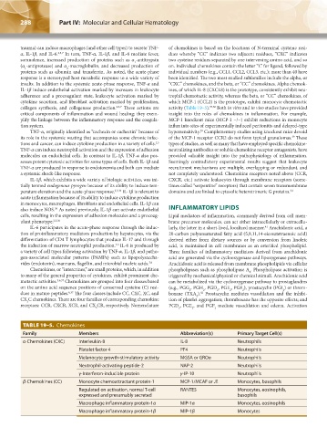
TABLE 19–5. Chemokines
Family Members Abbreviation(s) Primary Target Cell(s)
α-Chemokines (CXC) Interleukin-8 IL-8 Neutrophils
Platelet factor 4 PF4 Neutrophils
Melanocyte growth-stimulatory activity MGSA or GROα Neutrophils
Neutrophil-activating peptide-2 NAP-2 Neutrophils
γ-Interferon-inducible protein γ-IP-10 Neutrophils
β-Chemokines (CC) Monocyte chemoattractant protein-1 MCP-1/MCAF or JE Monocytes, basophils
Regulated on activation, normal T-cell RANTES Monocytes, eosinophils,
expressed and presumably secreted basophils
Macrophage inflammatory protein-1α MIP-1α Monocytes, eosinophils
Macrophage inflammatory protein-1β MIP-1β Monocytes
Kaushansky_chapter 19_p0279-0292.indd 288 9/17/15 5:51 PM