
Page 44 - Williams Hematology ( PDFDrive )
P. 44
20 Part I: Clinical Evaluation of the Patient Chapter 2: Examination of Blood Cells 21
MORPHOLOGIC EXAMINATION film interpretation. For example, leukemic blasts may appear dense
and rounded and lose their characteristic features when viewed in the
OF THE BLOOD thick part of the film. For specific purposes, the thick portion or side
and “feathered” edges of the film are of interest (for instance, to detect
Microscopic examination of the blood spread on a glass slide or cover- microfilariae and malarial parasites or to search for large abnormal cells
slip yields useful information regarding all the cells of the blood. The and platelet clumps).
process of preparing a thin blood film causes mechanical trauma to the The blood film is first scanned at low magnification (×200) to confirm
cells, introducing artifacts that can be minimized by good technique. reasonably even distribution of leukocytes and to check for abnormally
The optimal part of the stained blood film to use for morphologic large or immature cells in the side and feathered edges of the film. The
examination of the blood cells should be sufficiently thin that a small feathered edge is examined for platelet clumps. Abnormal cells, red cell
proportion of erythrocytes in a ×1000 magnification field touch each aggregation or rouleaux, background bluish staining consistent with para-
other, but not so thin that no red cells are touching. Figure 2–4 is a proteinemia, and parasites are all findings that can be suggested by medium
composite image taken from the optimal portion of the film showing magnification examination (×400). The optimal portion of the film is then
the five major leukocyte types, normal red cells, and platelets. Selection examined at high magnification (×1000, oil immersion) to systematically
of a portion of the blood film for analysis that is too thick or too thin assess the size, shape, and morphology of the major cell lineages.
for proper morphologic evaluation is the most common error in blood
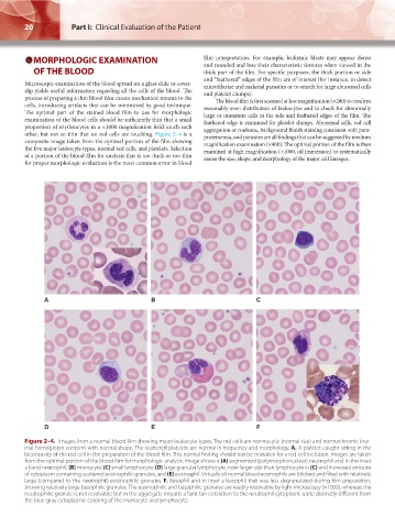
A B C
D E F
Figure 2–4. Images from a normal blood film showing major leukocyte types. The red cells are normocytic (normal size) and normochromic (nor-
mal hemoglobin content) with normal shape. The scattered platelets are normal in frequency and morphology. A. A platelet caught sitting in the
biconcavity of the red cell in the preparation of the blood film. This normal finding should not be mistaken for a red cell inclusion. Images are taken
from the optimal portion of the blood film for morphologic analysis. Image shows a (A) segmented (polymorphonuclear) neutrophil and in the inset
a band neutrophil; (B) monocyte; (C) small lymphocyte; (D) large granular lymphocyte, note larger size than lymphocyte in (C) and increased amount
of cytoplasm containing scattered eosinophilic granules; and (E) eosinophil. Virtually all normal blood eosinophils are bilobed and filled with relatively
large (compared to the neutrophil) eosinophilic granules. F. Basophil and in inset a basophil that was less degranulated during film preparation,
showing relatively large basophilic granules. The eosinophilic and basophilic granules are readily resolvable by light microscopy (×1000), whereas the
neutrophilic granule is not resolvable but in the aggregate imparts a faint tan coloration to the neutrophil cytoplasm, quite distinctly different from
the blue-gray cytoplasmic coloring of the monocyte and lymphocyte.
Kaushansky_chapter 02_p0011-0026.indd 20 17/09/15 5:35 pm