
Page 45 - Williams Hematology ( PDFDrive )
P. 45
20 Part I: Clinical Evaluation of the Patient Chapter 2: Examination of Blood Cells 21
RED CELL MORPHOLOGY densely stained and appear smaller because of their rounded shape, and
Normal erythrocytes on dried films are nearly uniform in size, with a show decreased or absent central pallor. A red cell with a spot or disc
mean diameter of approximately 7.5 μm (normal and abnormal red cells of hemoglobin within the central pale area is a target cell, in reality a
are described in more detail in Chap. 31). The normal-sized erythrocyte cup-shaped cell that distorts as it is flattened on the glass slide. These
is about the diameter of the nucleus of a small lymphocyte. The MCV is cells are typically found in disorders of hemoglobin synthesis (e.g., tha-
a more sensitive measure of red cell volume than the red cell diameter; lassemia), liver disease, and postsplenectomy where the cell-surface-to-
however, an experienced observer should be able to recognize abnor- cell-volume ratio is high. Chapter 31 describes the inclusions that may
malities in average red cell size when the MCV is significantly elevated be observed in erythrocytes on blood films.
or decreased. Anisocytosis is the term that describes variation in ery- Erythrocytes are usually distributed evenly throughout the blood
throcyte size, and is the morphologic correlate of the RDW. Macrocytes film. In some cases the cells become aligned in overlapping stacks,
and microcytes are red cells larger or smaller than normal, and their referred to as rouleaux (Chap. 109), resembling overlapping rows of
presence consistent with the measured MCV suggests certain diagnos- coins. Rouleaux are normal in the thick part of the film, but when found
tic possibilities. Early (“shift” or “stress”) reticulocytes (i.e., those with in the optimal viewing portion of the film, suggest a pathologic increase
the most residual RNA) appear in stained films as large, slightly blu- in immunoglobulin (Ig), particularly IgM-macroglobulinemia. Occa-
ish cells, referred to as polychromatophilic cells (Chap. 32). These cells sionally, high concentrations of IgA or IgG in patients with myeloma
are roughly the morphologic counterpart of the immature reticulocyte may also produce rouleaux.
fraction identified by automated instruments. The blood film is also useful to identify red cells with basophilic
The normal erythrocyte on a blood film is circular with central stippling (evidence of dyserythropoiesis), siderotic granules (evidence
pallor. Poikilocytosis is a term used to describe variations in the shape of sideroblastic erythropoiesis), Heinz bodies (evidence of unstable
of erythrocytes. The predominant appearance of a specific abnormality hemoglobins), and Howell-Jolly bodies (nuclear remnants). Microor-
in red cell shape can be an important diagnostic clue in patients with ganisms other than malaria parasites also may be found in or attached
anemia (Fig. 2–5). Erythrocytes with evenly spaced spikes (echinocytes to red cells (Chap. 53).
or crenated cells) can be an artifact caused by prolonged storage, or may
reflect metabolic erythrocyte abnormalities. PLATELET MORPHOLOGY
The normal erythrocyte appears as a disc with a rim of hemoglobin Platelets appear in normal stained blood film as small blue or colorless
and a clear central area, which normally occupies less than one-half the bodies with red or purple granules (see Fig. 2–4). Normal platelets aver-
cell diameter. Increased central pallor (hypochromia) is associated with age approximately 1 to 2 μm in diameter, but show wide variation in
disorders characterized by diminished hemoglobin synthesis, such as shape, from round to elongated, cigar-shaped forms. In improperly pre-
iron deficiency (Chap. 42). Evaluation of red cell hemoglobin content, pared films, platelets may form large aggregates in some areas and appear
as well as red cell size, is dependent on examining the proper part of to be diminished or absent in others. The frequent occurrence of giant
the blood film. Cells at the far “feathered edge” will always be large and platelets or platelet masses may indicate a myeloproliferative neoplasm
lack central pallor, whereas cells in the thick part of the film will look or improper collection of the blood specimen. The latter circumstance
small and rounded and will also lack central pallor. A sharp refractile can occur when venipuncture technique is faulty and platelets become
border demarcating the central area of pallor is an artifact secondary activated before the blood sample is thoroughly mixed with anticoag-
to inadequate drying of the film before staining (associated with high ulant. These platelet masses are apparent typically in the thin “feath-
humidity, and more common in anemic samples). Spherocytes are more ered edge” of the film, with corresponding fewer platelets elsewhere.
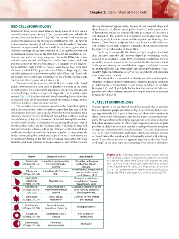
Figure 2–5. Disorders associated with certain red cell
Name Characteristic of Also seen in morphologic changes. Poikilocytosis is a general term used
Spherocyte Hereditary spherocytosis, Clostridial perfringens to indicate the presence of abnormally shaped red cells,
(Chaps. 31, 46, immune hemolytic septicemia, Wilson such as dacryocytes (teardrop-shaped red cells), schisto-
54) anemia disease cytes (fragmented red cells), and elliptocytes, as is found
Iron deficiency, megaloblastic in the most extreme form in hereditary pyropoikilocytosis
Elliptocyte Hereditary elliptocytosis anemia, thalassemia, (Chap. 46). MDS, myelodysplastic syndromes (Chap. 87).
(Chaps. 31, 46) (HE) myelofibrosis, MDS
Dacryocyte Severe iron deficiency,
(teardrop) Myelofibrosis megaloblastic anemia,
(Chaps. 31, 86) thalassemia, MDS
Schistocyte Microangiopathic, Occasional schistocytes are
(Chaps. 31, 51, mechanical hemolytic seen in many disorders
129, 132) anemia affecting red cells.
Echinocyte Renal failure, Common in vitro artifact
(Chaps. 31, 37) malnutrition after storage
Acanthocyte Spur cell anemia,
(Chaps. 31, 56) abetalipoproteinemia Postsplenectomy
Target cell Cholestasis, Hgb C Iron deficiency,
(Chaps. 31, 48) disease thalassemia
Stomatocyte Hereditary
(Chaps. 31, 46) stomatocytosis Alcoholism
Kaushansky_chapter 02_p0011-0026.indd 21 17/09/15 5:35 pm