
Page 46 - Williams Hematology ( PDFDrive )
P. 46
22 Part I: Clinical Evaluation of the Patient Chapter 2: Examination of Blood Cells 23
A B C
D E F
G H
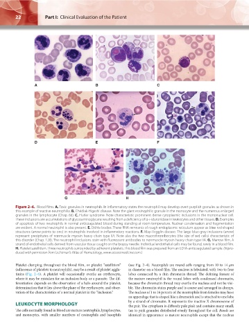
Figure 2–6. Blood films. A. Toxic granules in neutrophils. In inflammatory states the neutrophil may develop overt purplish granules as shown in
this example of reactive neutrophilia. B. Chédiak-Higashi disease. Note the giant eosinophilic granule in the monocyte and the numerous enlarged
granules in the lymphocyte (Chap. 66). C. Hurler syndrome. Note characteristic prominent dense cytoplasmic inclusions in the mononuclear cell.
These inclusions are accumulations of glycosaminoglycans resulting from a deficiency of α-l-iduronidase in leukocytes and other tissues. D. Examples
of apoptosis of two neutrophils in normal anticoagulated blood during standing at room temperature. Nuclear condensation and fragmentation
are evident. A normal neutrophil is also present. E. Döhle bodies. These RNA remnants of rough endoplasmic reticulum appear as blue rod-shaped
structures (arrow points to one) in neutrophils involved in inflammatory reactions. F. May-Hegglin disease. The large blue-gray inclusions (arrow)
represent precipitates of nonmuscle myosin heavy chain type IIA. Note also the two macrothrombocytes (the size of red cells) characteristic of
this disorder (Chap. 120). The neutrophil inclusions stain with fluorescent antibodies to nonmuscle myosin heavy chain type IIA. G. Marrow film. A
strand of endothelial cells derived from vascular tissue caught on the biopsy needle. Individual endothelial cells may be found, rarely in a blood film.
H. Platelet satellitism. Three neutrophils surrounded by adherent platelets. This blood film was prepared from an EDTA-anticoagulated sample. (Repro-
duced with permission from Lichtman’s Atlas of Hematology, www.accessmedicine.com.)
Platelet clumping throughout the blood film, or platelet “satellitism” (see Fig. 2–4). Neutrophils are round cells ranging from 10 to 14 μm
(adherence of platelets to neutrophils), may be a result of platelet agglu- in diameter on a blood film. The nucleus is lobulated, with two to four
tinins (Fig. 2–6). A platelet will occasionally overlie an erythrocyte, lobes connected by a thin chromatin thread. The defining feature of
where it may be mistaken for an inclusion body or a parasite. The dif- the mature neutrophil is the round lobes with condensed chromatin,
ferentiation depends on the observation of a halo around the platelet, because the chromatin thread may overlie the nucleus and not be visi-
determination that it lies above the plane of the erythrocyte, and obser- ble. The chromatin stains purple and is coarse and arranged in clumps.
vation of the characteristics of a normal platelet in the “inclusion.” The nucleus of 1 to 16 percent of the neutrophils from females may have
an appendage that is shaped like a drumstick and is attached to one lobe
by a strand of chromatin. It represents the inactive X chromosome of
LEUKOCYTE MORPHOLOGY the pair. The cytoplasm is diffusely pale pink and contains many small,
The cells normally found in blood are mature neutrophils, lymphocytes, tan to pink granules distributed evenly throughout the cell. Bands are
and monocytes, with smaller numbers of eosinophils and basophils identical in appearance to mature neutrophils except that the nucleus
Kaushansky_chapter 02_p0011-0026.indd 22 17/09/15 5:35 pm