
Page 487 - Williams Hematology ( PDFDrive )
P. 487
462 Part VI: The Erythrocyte Chapter 31: Structure and Composition of the Erythrocyte 463
erythron for most of fetal life. In later fetal life, skeletal development differentiating erythroid cells (Fig. 31–1A). A number of binding pro-
provides marrow niches to which erythropoiesis relocates being sus- teins are implicated in the cell–cell adhesions important to this process.
tained in the form of erythroblastic islands, a central macrophage with These include α β integrin, erythroblast macrophage protein (EMP),
4 1
12
circumferential layers of developing erythroid cells. The definitive and intercellular adhesion molecule-4 (ICAM-4) on the erythroblasts
stage of erythroid maturation predominates during the remainder of and vascular cell adhesion molecule (VCAM-1), EMP, α integrin on
V
16
fetal development and is the only type of erythroid maturation present macrophages. Additional macrophage receptors include CD69 (sialo-
through childhood and adult life. All of normal human erythropoiesis adhesin) and CD163, but the counterreceptors for these on erythrob-
occurs in the marrow in the form of erythroblastic islands. 13 lasts remains to be defined. Phase-contrast microcinematography
16
reveals that the macrophage is far from passive or immobile. Evidence
ERYTHROID PROGENITORS suggests that either the erythroblastic islands migrate or that erythroid
Burst-Forming Unit–Erythroid precursors move from island to island, as islands near sinusoids are
The earliest identifiable progenitor committed to the erythroid lineage composed of more mature erythroblasts while islands more distant
18
is the BFU-E (see Chap. 32, Fig. 32–1). A BFU-E is defined in vitro by from the sinusoids are composed of proerythroblasts. The macro-
its ability to create a “burst” on semisolid medium—that is, a colony phage’s pseudopodium-like cytoplasmic extensions move rapidly over
consisting of several hundred to thousands of cells by 10 to 14 days of cell surfaces of the surrounding wreath of erythroblasts. On phase con-
growth, during which time smaller satellite clusters of cells form around trast micrographs, the central macrophage of the erythroblastic island
a larger central group of erythroid cells, giving rise to the designation of a appears sponge-like, with surface invaginations in which the erythrob-
“burst.” The generation of BFU-E from hematopoietic stem cells requires lasts lie (Fig. 31–1B). As the erythroblast matures, it moves along a
interleukin (IL)-3, stem cell factor, and erythropoietin for differentiation, cytoplasmic extension of the macrophage away from the main body.
proliferation, prevention of apoptosis, and maturation (Chap. 18). 5,13 When the erythroblast is sufficiently mature for nuclear expulsion, the
erythroblast makes contact with an endothelial cell, passes through a
Colony-Forming Unit–Erythroid pore in the cytoplasm of the endothelial cell and enters the circulation
As erythroid maturation progresses, a later progenitor, the CFU-E, as a polychromatophilic macrocyte (reticulocyte). 19–21 The nucleus is
derived from the BFU-E, can be defined in vitro. The CFU-E is depen- ejected prior to egress from the marrow, phagocytized, and degraded
22
dent on erythropoietin for its development and can undergo only a few by marrow macrophages. In addition to the unique cytologic features
cell divisions. Thus, the CFU-E forms a smaller colony of morpho- described above, the macrophage of the erythroblastic island is also
5,14
logically recognizable erythroid cells in 5 to 7 days (see Chap. 32, Fig. molecularly distinct as demonstrated by a unique immunophenotypic
23
32–1). Adhesion between erythroid cells and macrophages occurs at the signature. In addition, the macrophage of the erythroblastic island
CFU-E stage of maturation. appears to play a stimulatory role in erythropoiesis independent of ery-
Using cell-surface markers, IL-3 receptor, CD34, and CD36, highly thropoietin. The anemia of chronic inflammation and of the myelodys-
purified populations of BFU-E and CFU-E can be isolated from human plastic syndrome (MDS) may result, at least in part, from inadequate
marrow. Gene expression profiling show distinctive changes in gene stimulation of erythropoiesis by these macrophages (Chap. 5).
5
expression profiles in hematopoietic stem cells, BFU-E, and CFU-E. Despite the central role of erythroid islands in erythropoiesis in
5
Some of the marrow failure syndromes are the result of defects in differ- vivo, morphologically normal development of erythroid cells can be
entiation of stem cells into erythroid progenitors. recapitulated in vitro without these structures as long as developing
cells are provided with supraphysiologic concentrations of appropriate
ERYTHROBLASTIC ISLAND cytokines and growth factors. Such growth, however, occurs at a much
slower rate than that observed in vivo, when erythroblasts form ery-
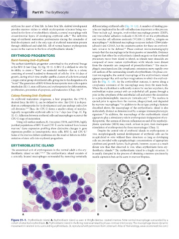
The anatomical unit of erythropoiesis in the normal adult is the ery- throblastic islands. The erythroblastic island is a fragile structure. It
24
throblastic island or islet. 13,16,17 The erythroblastic island consists of is usually disrupted in the process of obtaining a marrow specimen by
a centrally located macrophages surrounded by maturing terminally needle aspiration but can be seem in marrow biopsies.
A B
Figure 31–1. Erythroblastic island. A. Erythroblastic island as seen in Wright-Giemsa–stained marrow. Note central macrophage surrounded by a
cohort of attached erythroblasts. B. Erythroblastic island in the living state examined by phase-contrast microscopy. The macrophage shows dynamic
movement in relation to its surrounding erythroblasts. (A, reproduced with permission from Lichtman’s Atlas of Hematology, www.accessmedicine.com.)
Kaushansky_chapter 31_p0459-0478.indd 462 9/18/15 10:58 PM