
Page 492 - Williams Hematology ( PDFDrive )
P. 492
466 Part VI: The Erythrocyte Chapter 31: Structure and Composition of the Erythrocyte 467
may be twice the normal volume, with a corresponding increase in mean
cell hemoglobin (MCH) content. Whether the increase results from one
less mitotic division during maturation or from some other process such
as changes in cell cycle is not clear. It is interesting to note that mice
do not have the ability to produce stress reticulocytes with increased
mean cell volume (MCV) and MCH. In contrast, even under moderate
erythropoietic stress, some reticulocytes in the marrow pool shift to the
circulating pool. These “shift” reticulocytes with normal MCH contain
a higher-than-normal RNA content and now can be quantified. Quan-
tification is commonly performed by applying a fluorescent stain to tag
RNA and then dividing reticulocytes into high-, medium-, and low-fluo-
rescence categories using a fluorescence-sensitive flow cytometer. The
“stress” reticulocytes of the older literature likely fall in the high- and
medium-fluorescence categories. Unfortunately, at present little atten-
tion is being paid to discriminate stress and shift reticulocytes.
Pathology of the Reticulocyte
The reticulocyte may show pathologic alterations in size or staining
properties. The reticulocyte may contain inclusions visible by light
microscopy or identifiable only on ultrastructural analysis. Most patho-
logic inclusions usually attributed to erythrocytes are actually found
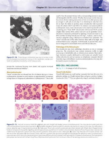
Figure 31–10. Orthochromic erythroblast ejecting its nucleus. A thin within reticulocytes and are nuclear or cytoplasmic remnants derived
rim of cytoplasm surrounds the nucleus. In the cytoplasm, a single cen- from late-stage erythroblasts. In splenectomized patients, they may also
triole (c) is partially encircled by some Golgi saccules. be found in mature erythrocytes.
RED CELL INCLUSIONS
process the membrane becomes more elastic and acquires increased
membrane mechanical stability. 32 See Fig. 31–11 for images of red cell inclusions.
Macroreticulocytes Howell-Jolly Bodies
“Stress” reticulocytes are released into the circulation during an intense Howell-Jolly bodies are small nuclear remnants that have the color of a
erythropoietin response to acute anemia or experimentally in response pyknotic nucleus on Wright-stained films and give a positive Feulgen
to large doses of exogenously administered erythropoietin. These cells reaction for DNA. 35,36 They are spherically shaped, randomly distributed
34
A B C
D E F
Figure 31–11. Red cell inclusions. Blood films. A. Red cells with Howell-Jolly bodies (arrows) postsplenectomy. The crisp circular border, dark blue
color, and peripheral location are characteristic. B. Basophilic stippling. These basophilic inclusions may be fine or coarse. In this case, the cell contains
coarse stippling seen in lead poisoning (arrow). C. Siderocyte. These cells contain purple granules when stained with Wright stain (Pappenheimer
bodies). Compared to basophilic stippling, siderotic granules are usually fewer in number and sometimes clustered. These Prussian blue–stained cells
confirm that the granules contain iron (blue reaction product). Arrow points to two siderocytes. D. Cabot ring. Rare red cell inclusion (arrow). See
text for further description. E. Heinz bodies. These cells from a patient with glucose-6-phosphate dehydrogenase deficiency were incubated with a
supravital dye, which stains the denatured globin precipitates. F. Red cells from a patient with hemoglobin H disease (α-thalassemia). The hemoglobin
precipitates are stained with brilliant cresyl blue. (Reproduced with permission from Lichtman’s Atlas of Hematology, www.accessmedicine.com.)
Kaushansky_chapter 31_p0459-0478.indd 467 9/18/15 10:59 PM