
Page 489 - Williams Hematology ( PDFDrive )
P. 489
464 Part VI: The Erythrocyte Chapter 31: Structure and Composition of the Erythrocyte 465
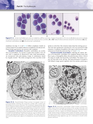
A B C D
Figure 31–3. Human erythroblast precursors as isolated by cell flow cytometry. Images of populations of human erythroblast precursors at stages
of erythroid maturation when sorted from human marrow by flow cytometry. A and B. Proerythroblasts and early basophilic erythroblasts; (C) poly-
chromatic erythroblasts; and (D) orthochromatic erythroblasts.
membrane (see Figs. 31–2 and 31–4) Diffuse cytoplasmic density on spokes or a clock face. The cytoplasm stains deep blue, leaving a perinu-
sections stained for peroxidase indicates hemoglobin is already present. clear halo that expands into a juxtanuclear clear zone around the Golgi
Dispersed glycogen particles are present in the cytoplasm. apparatus. Cytoplasmic basophilia at this stage results from the contin-
Basophilic Erythroblasts Basophilic erythroblasts are smaller ued presence of polyribosomes (see Figs. 31–2 and 31–5).
than proerythroblasts. The nucleus occupies three-fourths of the cell Polychromatophilic Erythroblasts Following the mitotic divi-
area and is composed of characteristic dark violet heterochroma- sion of the basophilic erythroblast, the cytoplasm changes from deep
tin interspersed with pink-staining clumps of euchromatin linked blue to gray, as hemoglobin dilutes the polyribosome content. Cells at
by irregular strands. The whole arrangement often resembles wheel this stage are smaller than basophilic erythroblasts. The nucleus occu-
13
pies less than half of the cell area. The heterochromatin is located in
well-defined clumps spaced regularly about the nucleus, producing a
n pr
p
g
g
n
Figure 31–4. Proerythroblast. Phase-contrast micrograph (inset) of pr
a proerythroblast showing the immature nucleus with nucleoli and
finely dispersed nuclear chromatin. The centrosome (juxtanuclear clear
zone) is apparent with its dense accumulation of mitochondria. Electron Figure 31–5. Basophilic erythroblast. Phase-contrast photomicro-
microscopic section of the proerythroblast shows nucleoli (n) in contact graph (inset) shows increased clumping of the nuclear chromatin and
with the nuclear membrane. Chromatin is finely dispersed and forms further rounding of the cell, with aggregation of the mitochondria and
small aggregates in the fixed nuclear membrane. The perinuclear canal centrosome into the regions of nuclear indentation. Electron micro-
is narrow but well defined. Polyribosome groups, many in helical config- scopic section shows clumping of the nuclear chromatin, nuclear pores
uration, are dispersed throughout the cytoplasm. The Golgi apparatus (p), organization of the nucleoli, increased density of polyribosomes (pr),
(g) is well developed, and regions of endoplasmic reticulum (arrows) are well-developed Golgi apparatus (g), and a decrease in smooth endo-
seen. plasmic reticulum.
Kaushansky_chapter 31_p0459-0478.indd 464 9/18/15 10:58 PM