
Page 490 - Williams Hematology ( PDFDrive )
P. 490
464 Part VI: The Erythrocyte Chapter 31: Structure and Composition of the Erythrocyte 465
C
pnc
P
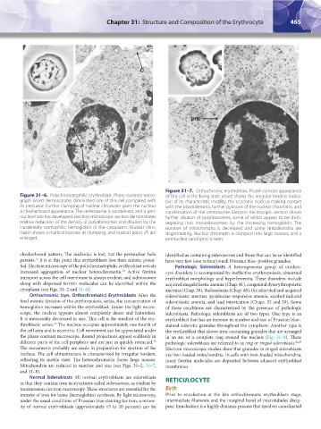
Figure 31–7. Orthochromic erythroblast. Phase-contrast appearance
Figure 31–6. Polychromatophilic erythroblast. Phase-contrast micro- of this cell in the living state (inset) shows the irregular borders indica-
graph (inset) demonstrates diminished size of this cell compared with tive of its characteristic motility, the eccentric nucleus making contact
its precursor. Further clumping of nuclear chromatin gives the nucleus with the plasmalemma, further pyknosis of the nuclear chromatin, and
a checkerboard appearance. The centrosome is condensed, and a peri- condensation of the centrosome. Electron microscopic section shows
nuclear halo has developed. Electron microscopic section demonstrates further dilution of polyribosomes, some of which appear to be disin-
relative reduction of the density of polyribosomes and dilution by the tegrating into monoribosomes, by the increasing hemoglobin. The
moderately osmiophilic hemoglobin in the cytoplasm. Nuclear chro- number of mitochondria is decreased, and some mitochondria are
matin shows a marked increase in clumping, and nuclear pores (P) are degenerating. Nuclear chromatin is clumped into large masses, and a
enlarged. perinuclear canal (pnc) is seen.
checkerboard pattern. The nucleolus is lost, but the perinuclear halo identified as containing siderosomes and those that can be so identified
persists. It is at this point that erythroblasts lose their mitotic poten- have very few (one to four) small Prussian blue–positive granules.
13
tial. Electron microscopy of the polychromatophilic erythroblast reveals Pathologic Sideroblasts A heterogeneous group of erythro-
13
increased aggregation of nuclear heterochromatin. Active ferritin cyte disorders is accompanied by ineffective erythropoiesis, abnormal
transport across the cell membrane is always evident, and siderosomes erythroblast morphology and hyperferremia. These disorders include
along with dispersed ferritin molecules can be identified within the acquired megaloblastic anemia (Chap. 41), congenital dyserythropoietic
cytoplasm (see Figs. 31–2 and 31–6). anemias (Chap. 39), thalassemias (Chap. 48), the inherited and acquired
Orthochromic (syn. Orthochromatic) Erythroblasts After the sideroblastic anemias, pyridoxine-responsive anemia, alcohol-induced
final mitotic division of the erythropoietic series, the concentration of sideroblastic anemia, and lead intoxication (Chaps. 52 and 59). Some
hemoglobin increases within the erythroblast. Under the light micro- of these conditions are characterized by the presence of pathologic
scope, the nucleus appears almost completely dense and featureless. sideroblasts. Pathologic sideroblasts are of two types. One type is an
It is measurably decreased in size. This cell is the smallest of the ery- erythroblast that has an increase in number and size of Prussian blue–
13
throblastic series. The nucleus occupies approximately one-fourth of stained siderotic granules throughout the cytoplasm. Another type is
the cell area and is eccentric. Cell movement can be appreciated under the erythroblast that shows iron-containing granules that are arranged
the phase-contrast microscope. Round projections appear suddenly in in an arc or a complete ring around the nucleus (Fig. 31–8). These
different parts of the cell periphery and are just as quickly retracted. pathologic sideroblasts are referred to as ring or ringed sideroblasts. 26,27
13
The movements probably are made in preparation for ejection of the Electron microscopic studies show that granules in ringed sideroblasts
nucleus. The cell ultrastructure is characterized by irregular borders, are iron-loaded mitochondria. In cells with iron-loaded mitochondria,
reflecting its motile state. The heterochromatin forms large masses. many ferritin molecules are deposited between adjacent erythroblast
Mitochondria are reduced in number and size (see Figs. 31–2, 31–7, membranes.
and 31–8).
Normal Sideroblasts All normal erythroblasts are sideroblasts
in that they contain iron in structures called siderosomes, as evident by RETICULOCYTE
transmission electron microscopy. These structures are essential for the Birth
transfer of iron for heme (hemoglobin) synthesis. By light microscopy, Prior to enucleation at the late orthochromatic erythroblasts stage,
under the usual conditions of Prussian blue staining for iron, a minor- intermediate filaments and the marginal band of microtubules disap-
ity of normal erythroblasts (approximately 15 to 20 percent) can be pear. Enucleation is a highly dynamic process that involves coordinated
Kaushansky_chapter 31_p0459-0478.indd 465 9/18/15 10:58 PM