
Page 491 - Williams Hematology ( PDFDrive )
P. 491
466 Part VI: The Erythrocyte Chapter 31: Structure and Composition of the Erythrocyte 467
The smaller portion consists of the expelled nucleus surrounded by a
thin ring of hemoglobin and plasma membrane (Fig. 31–9). In vivo,
expulsion of the nucleus may occur while the erythroblast is still part
of an erythroblastic island or the nucleus may be lost during passage
through the wall of a marrow sinus as the nucleus, which cannot tra-
verse the small opening, remains in the marrow. The outer leaflet of
the bilaminar membrane surrounding the expelled nucleus is high in
22
phosphatidylserine, a signal for macrophage ingestion (Fig. 31–10). It
is not clear what fraction of the expelled nuclei is ingested by the mac-
rophage of the erythroblastic island or by other macrophages resident in
marrow. Two hypotheses have been proposed to explain how the retic-
ulocyte exits the marrow. 19–21 The reticulocyte may actively traverse the
sinus epithelium to enter the lumen. More likely, however, the reticulo-
cyte may be driven across by a pressure differential because it appears
incapable of directed amoeboid motion. The precise mechanism is yet
to be defined.
Maturation
Following nuclear extrusion, the reticulocyte retains mitochondria,
small numbers of ribosomes, the centriole, and remnants of the Golgi
apparatus. It contains no endoplasmic reticulum. Supravital staining
with brilliant cresyl blue or new methylene blue produces aggregates
of ribosomes, mitochondria, and other cytoplasmic organelles. These
aggregates stain deep blue and, arranged in reticular strands, give the
reticulocyte its name. Maturation of the reticulocyte requires 48 to
72 hours. During this period, approximately 20 percent of the membrane
surface area is lost and cell volume decreases by 10 to 15 percent and the
final assembly of the membrane skeleton is completed. 31–33 Living retic-
ulocytes observed by phase-contrast microscopy are irregularly shaped
cells with a characteristically puckered exterior and a motile membrane.
Figure 31–8. Pathologic sideroblast is an erythroblast characterized
by the presence of mitochondrial deposits of iron-containing ferrugi- Examined by electron microscopy, reticulocytes are irregularly shaped
13
nous micelles (arrows) between the cristae. and contain many remnant organelles. The organelles, small smooth
vesicles, and an occasional centriole are grouped in the region of the cell
where the nucleus is expelled. In “young” reticulocytes, the vast major-
action of multiple mechanisms. 28–30 Tubulin and actin become concen- ity of ribosomes dispersed throughout the cytoplasm are in the form of
trated at the point where the nucleus will exit. These changes, accom- polyribosomes. As protein synthesis diminishes during maturation, the
panied by microtubular rearrangements and actin polymerization, play polyribosomes gradually transform into monoribosomes. During retic-
a role in nuclear expulsion. Expulsion of the nucleus in vitro is not an ulocyte maturation there is significant remodeling of the membrane,
instantaneous phenomenon; it requires a period of 6 to 8 minutes. The including loss of membrane proteins that include transferrin receptors,
process begins with several vigorous contractions around the midpor- Na-K adenosine triphosphatase (ATPase), and adhesion molecules, as
tion of the cell, followed by a division of the cell into unequal portions. well as loss of tubulin and cytoplasmic actin. During the remodeling
33
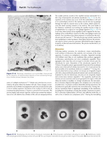
A B C D
Figure 31–9. Morphology of cells during reticulocyte maturation. A. Orthochromatic erythroblast extruding its nucleus. B. Multilobular, motile
reticulocyte generated following nuclear extrusion. C. The cup-shaped, nonmotile reticulocyte at a later stage of maturation. D. Mature discoid red cell.
Kaushansky_chapter 31_p0459-0478.indd 466 9/18/15 10:58 PM