
Page 593 - Williams Hematology ( PDFDrive )
P. 593
568 Part VI: The Erythrocyte Chapter 39: The Congenital Dyserythropoietic Anemias 569
Allogeneic marrow transplantation from an human leukocyte OTHER CONGENITAL
antigen (HLA)-identical sibling has been successful in transfusion-
dependent children with very severe CDA II and in one adult with CDA DYSERYTHROPOIETIC ANEMIAS
II and β-thalassemia trait. 32,33,55,56
A number of cases of CDA that do not have specific features of types I,
II, or, to some extent, III disease have been reported. 69–74 Classification
75
CONGENITAL DYSERYTHROPOIETIC has been proposed based largely on cell morphology. In addition, sev-
eral genes have been associated with CDA variants, including mutations
ANEMIA, TYPE III in the GATA1 and KLF1 genes, which are critical for development of
specific blood cell lineages (outlined in Table 39–1). 69–74,76
CLINICAL AND LABORATORY FINDINGS Alterations in the erythroid hematopoietic transcription factor
77
Type III is the least-common form of CDA. This condition, which is KLF1 gene are associated with CDA type IV, characterized by severe
dominantly inherited, was initially coined “hereditary benign erythro- hemolytic anemia, elevated fetal hemoglobin, and deficiency of ery-
cytosis.” One dominantly inherited form was reported as early as 1951 throid proteins CD44 and aquaporin 1 (see Table 39–1). 78
in a woman and her three children, in whom 16.0 to 22.7 percent of Additionally, syndromes have been described in which CDA
hematopoietic stem cell erythroblasts were multinucleated. Giant-size accounts only for one feature of a syndrome phenotype (see Table 39–1).
erythrocytes were present in the blood. For instance, Majeed syndrome, is comprised of chronic recurrent mul-
Most of our knowledge about CDA III stems from a large family tifocal osteomyelitis, inflammatory dermatosis, and CDA. The respon-
from the province of Västerbotten in northern Sweden. The diagnosis sible gene is LPIN2 (18p11.31), encoding lipin 2, an ER-phosphatidate
57
79
was made in the adults and older children. The spleen was not palpable, phosphatase. Additionally, dyserythropoiesis associated with exo-
nor was iron overload recorded. The large size of this family made it crine pancreatic insufficiency and calvarial hyperostosis resulting from
80
possible to map the responsible gene to 15q22–25. In addition, a num- COX4I2 gene mutations has been described in two Arab families.
58
ber of sporadic cases of CDA III have been reported. 59 Mevalonate kinase deficiency (MKD) resulting from a missense muta-
In another case, a number of stillbirths, including at least one tion in the MVK gene and showing morphologic marrow cell abnormal-
stillborn with hydrops fetalis, were noted in an Indian family in which ities similar to CDA II has also been reported. 81
the mother, who initially required transfusions, became transfu-
sion-independent after splenectomy. 60 DIFFERENTIAL DIAGNOSIS
Blood films from these patients show macrocytes, occasional
extremely large forms (gigantocytes), and poikilocytes. Patients are Congenital dyserythropoietic anemias may be confused with thalas-
generally asymptomatic, with no or moderate anemia, mild jaundice, semias and other hemolytic anemias. Marked anisocytosis, including a
and, commonly, cholelithiasis. The reticulocyte count is typically less
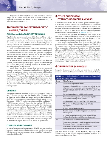
than 3 percent. 57,61 Marrow shows marked erythroid hyperplasia, with TABLE 39–1. A Classification Frame for Atypical Congenital
large multinucleate erythroblasts with large, lobulated nuclei, and giant Dyserythropoietic Anemias
multinucleate erythroblasts with up to 12 nuclei (see Fig. 39–2A). On
electron microscopy, clefts within heterochromatin, autophagic vacu- Group Main Features
oles, iron-laden mitochondria, and myelin figures in the cytoplasm have IV Transfusion-dependent anemia
been reported. 62 Pronounced normoblastic erythroid hyperplasia with a
slight to moderate increase in the nonspecific dysery-
GENETICS thropoietic erythroblasts with irregular or karyorrhectic
nuclei
The causative mutation was found to be 2747C>G (P916R) in the KIF23 No precipitated protein within erythroblasts
63
gene. The same mutation was also found in CDA III patients from
an American family without any known relation to the Swedish kin- V Near-normal hemoglobin with normal or slightly
dred. KIF23 encodes a kinesin-superfamily molecule, mitotic kinesin- increased mean corpuscular volume
63
like protein 1 (MKLP1), a mitotic protein essential for cytokinesis. 64,65 Predominantly unconjugated hyperbilirubinemia
MKLP1 interacts with Arf6 (adenosine diphosphate (ADP)-ribosy- Marked normoblastic/slightly megaloblastic
lation factor 6), ultimately forming an extended β-sheet that interacts hyperplasia
with the membrane surface at the cleavage furrow. The Arf6–MKLP1
complex plays a crucial role in cytokinesis by connecting the microtu- Little or no erythroid dysplasia
bule bundle and membranes at the cleavage plane. Knockdown of Arf6 VI Normal or near-normal hemoglobin with marked
64
results in binucleated and multinucleated cells, a hallmark of CDA III macrocytosis
erythroblasts. In knockdown and rescue experiments with in vitro cell Erythroid hyperplasia with cobalamin- and folate-
66
lines, cytokinesis failure and binucleated cells were seen more frequently independent florid megaloblastic erythropoiesis
with the P916R mutant than wild-type GFP-MKLP1, indicating that the
P916R mutation impairs the function of MKLP1 in cytokinesis. 63 VII Severe transfusion-dependent anemia
Severe normoblastic erythroid hyperplasia with irregu-
lar nuclear shapes in many erythroblasts
COURSE AND PROGNOSIS Intraerythroblastic inclusions that resemble precipi-
In spite of an apparently benign course, CDA III is prone to various tated globin but do not contain globin
long-term complications, including intravascular hemolysis, increased
risk of myeloma and other monoclonal gammapathies, and develop- Data from Wickramasinghe SN and Wood WG: Advances in the
67
ment of angioid streaks. 68 understanding of the congenital dyserythropoietic anaemias. Br J
Haematol 131(4):431–446, 2005.
Kaushansky_chapter 39_p0563-0570.indd 568 9/17/15 6:21 PM