
Page 598 - Williams Hematology ( PDFDrive )
P. 598
572 Part VI: The Erythrocyte Chapter 40: Paroxysmal Nocturnal Hemoglobinuria 573
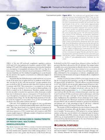
GPI-anchored protein Transmembrane protein Figure 40–2. The molecular and genetic basis of par-
oxysmal nocturnal hemoglobinuria (PNH). There are two
types of anchoring mechanisms for plasma membrane
proteins: transmembrane and glycosylphosphatidylinosi-
Protein tol (GPI). Transmembrane proteins are anchored into the
lipid bilayer of the cell by a short series (approximately
25 amino acids) of hydrophobic residues (blue rectangle).
EtN
Man EtN Transmembrane proteins typically have a short cytoplas-
Man mic tail that usually has signaling properties (red rectan-
Man gle). The ectoplasmic portion of the protein is illustrated by
EtN PNH defect the series of gray-blue squares. The GPI-anchored protein
GlcN (AP) consists of the following components: phosphati-
dylinositol (inositol is represented by the blue hexagon
labeled I and phosphate is represented by the red oval);
I glucosamine (GLcN, yellow circle); three mannose (Man,
green circles); ethanolamine phosphate (EtN, blue square
with attached phosphate represented by the red oval); the
protein entity (blue circle). The lipid component (indicated
by the series of diagonal lines within the lipid bilayer) is
usually 1-alkyl,2-acylglycerol for mammalian GPI-APs. PNH
Lipid bilayer cells are deficient in all GPI-APs because somatic mutation
of the X-chromosome gene PIGA disrupts the first step in
the biosynthetic pathway (transfer of the nucleotide sugar
UDP-GlcNAc [uridine diphosphate–N-acetylglucosamine]
to GlcNAc-PI [phosphatidylinositol]) indicated by the
arrow.
(RBCs) of the two GPI-anchored complement regulatory proteins, be derived from the PIGA-mutant clone, whereas in others, less than 10
CD55 and CD59, that underlies the hemolytic anemia of PNH. RBCs percent of the blood cells may be GPI-AP deficient. This unique feature
17
lacking CD55 and CD59 undergo spontaneous intravascular hemolysis (variability in extent of mosaicism) is clinically relevant because patients
as a consequence of unregulated activation of the APC (see Fig. 40–1, with relatively small PNH clones have minimal or no symptoms and
bottom panel). Thus, the hallmark clinical manifestation of PNH (intra- require no PNH-specific treatment, whereas those with large clones are
vascular hemolysis and the resultant hemoglobinuria) occurs because often debilitated by the consequences of chronic complement-medi-
the two proteins that regulate complement on erythrocytes happen to ated intravascular hemolysis and respond dramatically to complement
be GPI-anchored. inhibitory therapy.
Hypothetically, the PNH phenotype would result from inactivation Another remarkable feature of PNH is phenotypic mosaicism (see
of any of the more than 25 genes involved in synthesis of the GPI-an- Fig. 40–3A) based on PIGA genotype (see Fig. 40–3B) that determines
27
chor (see Fig. 40–2), but, with one exception, somatic mutation of no the degree of GPI-AP deficiency. PNH III cells are completely deficient
18
17
gene involved in GPI-AP synthesis other than PIGA has been reported in GPI-APs, PNH II cells are partially (approximately 90 percent) defi-
in patients with PNH. This phenomenon is accounted for by the fact cient and PNH I cells express GPI-APs at normal density (putatively,
that, of the genes involved in the GPI-anchor synthesis pathway, only these cells are progeny of residual normal stem cells; see Fig. 40–3A).
PIGA is located on the X-chromosome. Therefore, somatic mutation Phenotype varies among patients (Fig. 40–4). Some patients have only
of only one allele is required for expression of the phenotype as males type I and type III cells (the most common phenotype), some have type
have one X-chromosome and, as a consequence of X-inactivation dur- I, type II, and type III (the second most common phenotype), and some
ing embryogenesis, females have only one functional X-chromosome patients have only type I and type II cells (the least-common pheno-
in somatic tissues (Chap. 10). On the other hand, mutation of two alle- type). Furthermore, the contribution of each phenotype to the compo-
les would be required for inactivation of any of the autosomal genes sition of the blood varies. Phenotypic mosaicism is clinically important
involved in the GPI-anchor synthesis pathway. 18 because PNH II cells are relatively resistant to spontaneous hemolysis,
Cells with PIGA mutations do not appear to have a proliferative and patients with a high percentage of type II cells have a relatively
advantage in vitro or in hybrid animal models made with PIGA knock- benign clinical course with respect to hemolysis (Fig. 40–4).
outs. They have been found to be relatively resistant to apoptosis in The anemia of PNH is multifactorial as an element of marrow fail-
19
some studies, 20–23 but not in others. 24,25 Thus, the basis of clonal selection ure is present in all patients, although the degree of marrow dysfunc-
and clonal expansion of PIGA mutant stem cells in patients with PNH tion is variable. In some patients, PNH arises in the setting of aplastic
28
remains largely enigmatic although a number of hypotheses have been anemia. In this case, marrow failure is the dominant cause of anemia.
26
proposed (reviewed in Ref. 17). In other patients with PNH, evidence of marrow dysfunction may be
subtle (e.g., an inappropriately low reticulocyte count) with the degree
PHENOTYPIC MOSAICISM IS CHARACTERISTIC of anemia being determined primarily by the rate of hemolysis that is,
OF PAROXYSMAL NOCTURNAL in turn, determined by PNH clone size.
HEMOGLOBINURIA CLINICAL FEATURES
The blood of patients with PNH is a mosaic of normal and abnormal
cells (Fig. 40–3). Although PNH is a clonal disease, the extent to which The primary clinical manifestations of PNH are hemolysis, thrombosis,
28
the PIGA-mutant clone expands varies widely among patients. As an and marrow failure. Constitutional symptoms (fatigue, lethargy, mal-
17
example, in some cases, greater than 90 percent of the blood cells may aise, asthenia) dominate the history, with nocturnal hemoglobinuria
Kaushansky_chapter 40_p0571-0582.indd 573 9/17/15 6:22 PM