
Page 597 - Williams Hematology ( PDFDrive )
P. 597
572 Part VI: The Erythrocyte Chapter 40: Paroxysmal Nocturnal Hemoglobinuria 573
cells, and the extent of the mosaicism varies widely among patients Alternative Pathway of Complement
(see “Phenotypic Mosaicism is Characteristic of Paroxysmal Nocturnal Eculizumab
Hemoglobinuria” below). Patients with small PNH clones have few or Membrane Attack Complex
no symptoms related to hemolysis. Thus an argument can be made that C3 convertase C3a C5 convertase C5 C5a
asymptomatic patients with small clones do not have clinically signifi- C3bBbP C3bBbC3bP C5b-9 n
cant PNH and should be excluded from prevalence estimates. Others,
however, may argue that any patient with flow cytometric evidence of CD55 CD55 CD59
a population of GPI-AP–deficient cells, regardless of clone size, has
PNH and should be included in prevalence estimates. Well-designed, Complement Activation
rigorous studies of prevalence that address the issue of disease het- LDH
erogeneity are needed, but, by any definition, PNH is a rare disease. LDH LDH
LDH
The prevalence of clinically significant PNH (i.e., classic PNH) plus LDH
patients with relatively large clones that arise in the setting of another LDH
marrow failure syndrome, (see “Clinical Features” and Table 40–2 Normal RBC PNH RBC
below) is likely in the order of less than 1 case per 200,000 population,
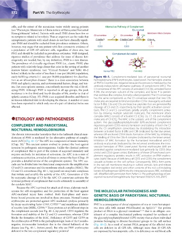
easily fulfilling criteria (<1 case per 50,000 population) for classifica- Figure 40–1. Complement-mediated lysis of paroxysmal nocturnal
5
tion as an ultraorphan disease. There is a close association between hemoglobinuria (PNH) erythrocytes. Upper panel. The hemolytic anemia
PNH and aplastic anemia, and environmental factors, drugs, and tox- of PNH is Coombs test–negative because the process is mediated by the
ins, that cause aplastic anemia, concordantly increase the risk of devel- antibody-independent alternative pathway of complement (APC). The
oping PNH. Although PNH is reported in all age groups, the peak C3 convertase of the APC consists of activated C3 (C3b), activated factor
incidence is in the third and fourth decades of life, similar to that of B (Bb, the enzymatic subunit of the complex), and factor P (a protein
that stabilizes the complex, formally called properdin). The C5 convertase
aplastic anemia (Chap. 35). PNH is an acquired disorder, and there is has the same components as the C3 convertase except that two C3b
no known inherited risk for developing the disease. A number of cases molecules are required to bind and position C5 for cleavage by activated
have been reported in which only one of a pair of identical twins was factor B (Bb). C3a and C5a are bioactive peptides that are generated by
affected. cleavage of C3 and C5, respectively, by their specific activation conver-
tases. The C3 and C5 convertases greatly amplify complement activa-
tion by cleaving multiple substrate molecules. The membrane attack
complex (MAC) consists of activated C5 (C5b), C6, C7, C8, and multiple
ETIOLOGY AND PATHOGENESIS molecules of C9 (C9 ). The MAC is the cytolytic unit of the complement
n
system. The glycosylphosphatidylinositol (GPI)-anchored complement
COMPLEMENT AND PAROXYSMAL regulatory protein CD55 restricts formation and stability of both the C3
NOCTURNAL HEMOGLOBINURIA and the C5 amplification convertases by destabilizing the interaction
between activated factor B (Bb) and C3b (indicated by the blue arrow),
The chronic intravascular hemolysis that is the hallmark clinical man- whereas GPI-anchored CD59 blocks formation of the MAC by inhibiting
ifestation of PNH is mediated by the alternative pathway of comple- the binding of C9 to the C5b-8 complex (indicated by the brown arrow).
ment (APC) (Fig. 40–1). The APC is a component of innate immunity Inhibition of MAC formation by the humanized monoclonal anti-C5
6
7
(Chap. 20). This ancient system evolved to protect the host against antibody eculizumab (indicated by the red arrow) ameliorates the intra-
invasion by pathogenic microorganisms. Unlike the classical pathway vascular hemolysis of PNH. Lower panel. Normal erythrocytes (left) are
of complement that is part of the system of acquired immunity and protected against complement-mediated lysis primarily by CD55 (blue
circles) and CD59 (green circles). Deficiency of these GPI-anchored com-
requires antibody for initiation of activation, the APC is in a state of plement regulatory proteins results in APC activation on PNH erythro-
continuous activation, armed at all times to protect the host (Chap. 19 cytes (right). Because of deficiency of CD55 and CD59, the complement
provides a detailed review of the complement system). The APC cas- cascade activates on the cell surface. Consequently, MACs form pores
cade can be divided into two functional components: the amplification in the red cell membrane resulting in colloid osmotic lysis and release
C3 and C5 convertases and the membrane attack complex (MAC). The of hemoglobin (red circles) and other contents of the red cell including
C3 and C5 convertases (Fig. 40–1, top panel) are enzymatic complexes lactate dehydrogenase (LDH) into the intravascular space. RBC, red blood
that initiate and amplify the activity of the APC. Generation of C5b cell. (Modified with permission from Parker CJ: The pathophysiology of par-
by enzymatic cleavage of C5 by the APC C5 convertase activates the oxysmal nocturnal hemoglobinuria. Exp Hematol 35(4):523–533, 2007.)
terminal pathway of complement that results ultimately in assembly of
the cytolytic MAC.
Because the APC is primed for attack at all times, elaborate mech-
anisms for self-recognition and for protection of the host against THE MOLECULAR PATHOGENESIS AND
APC-mediated injury have evolved. Both fluid-phase and mem- GENETIC BASIS OF PAROXYSMAL NOCTURNAL
brane-bound proteins are involved in these processes. Normal human
erythrocytes are protected against APC-mediated cytolysis primarily HEMOGLOBINURIA
by decay-accelerating factor (DAF, CD55) 8–10 and membrane inhibitor PNH is a consequence of clonal expansion of one or more hematopoi-
of reactive lysis (MIRL, CD59). These proteins act at different steps in etic stem cells with mutant PIGA(located on Xp22.1). The protein
11
12
the complement cascade (see Fig. 40–1, top panel). CD55 regulates the product of PIGA is a glycosyl transferase 12–16 that is an obligate con-
formation and stability of the C3 and C5 convertases, whereas CD59 stituent of a complex biochemical pathway required for synthesis of
blocks the formation of the MAC. Deficiency of CD55 and CD59 on the glycosylphosphatidylinositol (GPI) moiety that anchors individual
the erythrocytes of PNH is the pathophysiologic basis of the Coombs- proteins belonging to diverse functional groups to the cell surface
negative, intravascular hemolysis that is the clinical hallmark of the (Fig. 40–2). As a result of mutant PIGA, progeny of the affected stem
disease (see Fig. 40–1, bottom panel). But why are PNH erythrocytes cells are deficient in all GPI-APs. Although more than 20 GPI-APs
deficient in the two complement regulatory proteins? are expressed by hematopoietic cells, it is deficiency on red blood cells
Kaushansky_chapter 40_p0571-0582.indd 572 9/17/15 6:22 PM