
Page 653 - Williams Hematology ( PDFDrive )
P. 653
628 Part VI: The Erythrocyte Chapter 43: Iron Deficiency and Overload 629
IRON DEFICIENCY TABLE 43–1. Sources of Blood Loss
DEFINITION AND HISTORY ALIMENTARY TRACT
Iron deficiency is the state in which the content of iron in the body is Esophagus
less than normal. Iron depletion is the earliest stage of iron deficiency, in Varices
which storage iron is decreased or absent but serum iron concentration, Stomach and duodenum
transferrin saturation, and blood hemoglobin levels are normal. Ulcer
Iron deficiency without anemia is a somewhat more advanced stage of Hiatus hernia
iron deficiency, characterized by absent storage iron, usually low serum
iron concentration and transferrin saturation, but without frank anemia. Gastritis
Iron-deficiency anemia, the most advanced stage of iron deficiency, is Carcinoma
characterized by absent iron stores, low serum iron concentration, low Varices
transferrin saturation, and low blood hemoglobin concentration. Angiodysplasia
Chlorosis, or “green sickness,” was well known to European phy- Hemangioma
sicians after the middle of the 16th century. In France, by the middle
of the 17th century, iron salts and other remedies (including, oddly Leiomyoma (Ménétrier disease)
enough, phlebotomy) were used in its treatment. Not long thereafter, Mucosal hypertrophy
iron was recommended by Sydenham as a specific remedy for chlorosis. Hypergastrinemia
For the 100 years preceding 1930, iron was used in the treatment of Antral vascular ectasia
chlorosis, often in ineffective doses, although the mechanism of action “Watermelon stomach”
of iron and the appropriateness of its use were highly controversial. By
the beginning of the 20th century, it had been established that chlorosis Small intestine
was characterized by a decrease in the iron content of the blood and by Vascular ectasia
the presence of hypochromic erythrocytes, but it was not until the clas- Tumors
sic 1932 studies by Heath, Strauss, and Castle that it was shown that the Ulceration
1
response of anemia to iron was stoichiometrically related to the amount Meckel’s diverticulum
of iron given and that chlorosis was, indeed, iron deficiency. The history
of iron deficiency has been reviewed in greater detail elsewhere. 2,3 Colon and anorectal
Hemorrhoids
EPIDEMIOLOGY Carcinoma
Iron-deficiency anemia is the most common anemia worldwide, and Polyp
is especially prevalent in women and children in regions where meat Diverticulum
intake is low, food is not fortified with iron, and malaria, intestinal Ulcerative colitis
infections, and parasitic worms are common. Women with frequent Angiodysplasia
4–6
pregnancies may be particularly susceptible. In the United States, iron Hemangioma
deficiency is most common in children 1 to 4 years old and in adoles-
cent, reproductive age, or pregnant women. 7–9 Telangiectasia
Amebiasis
ETIOLOGY AND PATHOGENESIS BILIARY TRACT
Intrahepatic bleeding
Etiology
Iron deficiency may occur as a result of chronic blood loss, diversion Carcinoma
of iron to fetal and infant erythropoiesis during pregnancy and lacta- Cholelithiasis
tion, inadequate dietary iron intake, malabsorption of iron, intravascular Trauma
hemolysis with hemoglobinuria, diversion of iron to nonhematopoietic Ruptured aneurysm
tissues like the lung, genetic factors, or a combination of these factors. Aberrant pancreas
Of these, gastrointestinal or menstrual blood loss are the most common.
As discussed in Chap. 42, the average adult male has approximately 1000 GENITOURINARY TRACT
mg of iron in stores, but on average, women have less than half of this Menorrhagia
amount. The average daily dietary intake of iron is 10 to 12 mg, but much Uterine fibroids
of this is not absorbed, even when absorption is maximal. Blood loss Endometriosis
of each milliliter of packed erythrocytes represents 1 mg of iron. Thus Carcinoma
chronic daily blood loss greater than 5 mL of erythrocytes will deplete
iron reserves over weeks to months, and even if bleeding stops com- Vascular abnormalities
pletely, the repletion of lost iron, including the restoration of iron stores RESPIRATORY TRACT
(around 1000 mg in the average adult man), will take many months. Epistaxis
Carcinoma
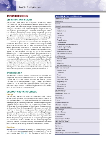
Blood Loss
Gastrointestinal Blood Loss In men and in postmenopausal women, Infections
iron deficiency is most commonly caused by chronic bleeding from the Telangiectases
gastrointestinal tract. Table 43–1 lists the causes of such blood loss. Idiopathic pulmonary hemosiderosis
Kaushansky_chapter 43_p0627-0650.indd 628 9/17/15 6:27 PM