
Page 656 - Williams Hematology ( PDFDrive )
P. 656
630 Part VI: The Erythrocyte Chapter 43: Iron Deficiency and Overload 631
Intravascular Hemolysis and Hemoglobinuria Normal Iron Iron- Iron-
Iron-deficiency anemia may occur in paroxysmal nocturnal hemo- depletion deficiency deficiency
globinuria (Chap. 40) and in hemolysis resulting from mechanical Storage anemia anemia
erythrocyte trauma from intracardiac myxomas, valvular prostheses iron (early) (advanced)
(particularly if malfunctioning), or patches (Chaps. 33 and 51). In these
disorders, up to 10 mg/day of iron is lost in the urine as hemosiderin
and ferritin in desquamated tubular cells and as hemoglobin dimers, an Hemoglobin
amount sufficient to cause systemic iron deficiency. 61,62 iron
Iron deficiency occurs frequently in athletes engaged in a variety
of sports (Chaps. 33 and 51), especially female athletes. There may be Transport
63
mild anemia. Increased intravascular hemolysis, presumably with some iron
renal loss of iron, may play a role, but gastrointestinal blood loss has also
been demonstrated in persons engaged in strenuous athletic pursuits. Enzyme
Hemoglobinuria and hemosiderinuria are also seen in competitive and iron
recreational runners, that is, march hemoglobinuria (Chaps. 33 and 51).
Strenuous exercise also elicits a rise in serum interleukin (IL)-6 and
hepcidin, and this could decrease dietary iron absorption. 63 Erthrocytes Normochromic Hypochromic
Women soldiers undergoing basic training experience iron deple- normocytic microcytic
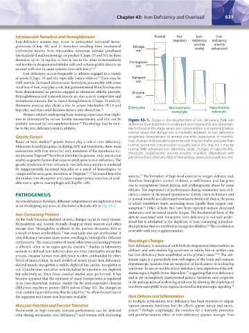
tion as determined by serum ferritin measurements, and this can be Figure 43–1. Stages in the development of iron deficiency. Early iron
partially reversed by iron supplementation. The etiology may be simi- deficiency (iron depletion) is usually not accompanied by any abnormali-
64
lar to the iron deficiency seen in athletes. ties in blood; at this stage, serum iron concentration is occasionally below
normal values and storage iron is markedly depleted. As iron deficiency
Genetic Factors progresses, development of anemia precedes appearance of morpho-
Based on twin studies, genetic factors play a role in iron deficiency. logic changes in blood, although some cells may be smaller and paler than
65
Mutations in multiple genes, including HFE and transferrin, show weak normal; serum iron concentration is usually low at this time, but it may be
associations with iron stores but only mutations of the membrane ser- normal. With advanced iron depletion, classic changes of hypochromic,
ine protease Tmprss6 have been identified in genome-wide association microcytic, hypoferremic anemia become manifest. (Reproduced with
66
permission from Lichtman’s Atlas of Hematology, www.accessmedicine.com.)
studies as genetic factors that cause or predispose to iron deficiency. The
genetic syndrome of iron-refractory iron deficiency anemia is mediated
by inappropriately increased hepcidin as a result of homozygous or
compound heterozygous mutations in Tmprss6. 67–69 Increased hepcidin 73
diminishes iron absorption and causes inappropriate retention of avail- anemia. The limitation of high-level exercise by oxygen delivery, and,
able iron in splenic macrophages and Kupffer cells. therefore, hemoglobin content of blood, is well known, and has given
rise to surreptitious blood doping and erythropoietin abuse by some
athletes. The impairment of performance during nonanemic iron defi-
PATHOGENESIS ciency consists of decreased spontaneous activity (seen in humans and
As iron deficiency develops, different compartments are depleted in iron in animal models) and decreased ventilatory threshold, that is, the point
at which ventilation starts increasing more rapidly than oxygen con-
in an overlapping sequence, as illustrated schematically in Fig. 43–1.
sumption. Other deficits that have been reported include decreased
74
endurance and increased muscle fatigue. The biochemical basis of the
Iron-Containing Proteins deficits associated with nonanemic iron deficiency is not well under-
As the body becomes depleted of iron, changes occur in many tissues. stood but is attributed to the depletion of iron-containing mitochon-
Hemosiderin and ferritin virtually disappear from marrow and other drial proteins that are involved in energy metabolism. The condition is
63
storage sites. Hemoglobin synthesis in the marrow decreases, first as reversible with iron supplementation.
a result of fewer erythroblasts, but eventually also per erythroblast if
70
iron deficiency becomes more severe, resulting in hemoglobin-deficient
erythrocytes. The concentration of many other iron-containing proteins Neurologic Changes
is affected, often in an organ-specific manner. Studies in laboratory Iron deficiency is associated with both developmental abnormalities in
71
animals on defined iron-deficient diets are most informative about this children and with restless leg syndrome in adults, but in neither case
process, because human iron deficiency is often confounded by other has iron deficiency been established as the primary cause. 75,76 The sub-
forms of malnutrition. In such models of severe (pure) iron deficiency, stantia nigra is a particularly iron-rich region of the brain and contains
skeletal muscle myoglobin is mildly depleted but cardiac myoglobin is dopaminergic neurons that are suspected of involvement in restless leg
not. Cytochromes and other mitochondrial ferroproteins are depleted syndrome. In mouse models of iron deficiency, iron depletion of the sub-
77
but selectively so. Since these classical studies were performed, it has stantia nigra is highly strain-dependent, suggesting that iron deficiency
become apparent that the synthesis of many ferroproteins is regulated and as yet incompletely characterized genetic variations may cooperate
in an iron-dependent manner, mainly via the iron-responsive element in the pathogenesis of restless leg syndrome by allowing the depletion of
(IRE)/iron-regulatory protein (IRP) system (Chap. 42). The changes in iron from susceptible brain regions involved in dopaminergic signaling. 78
iron-containing proteins may thus be adaptive, to allow the survival of
72
the organism until more iron becomes available. Host Defense and Inflammation
In multiple publications, iron deficiency has been reported to impair
Muscular Function and Exercise Tolerance various immune functions, but the effects appear minor and incon-
79
Decrements in high-intensity exercise performance can be detected sistent. Perhaps surprisingly, the evidence for a narrowly protective
even during nonanemic iron deficiency, and worsen with increasing and proinflammatory effect of iron deficiency appears stronger. Iron
63
Kaushansky_chapter 43_p0627-0650.indd 631 9/17/15 6:27 PM