
Page 658 - Williams Hematology ( PDFDrive )
P. 658
632 Part VI: The Erythrocyte Chapter 43: Iron Deficiency and Overload 633
(spoon nails), once a common finding, is now encountered rarely. Reti- abnormalities correct with iron therapy. Thrombotic complications of
nal hemorrhages and exudates may be seen in severely anemic patients iron deficiency have been reported but are very rare. The etiology
128
(e.g., hemoglobin concentration of <5 g/dL). Splenomegaly has occa- of either abnormality is not known. Low-iron-diet-induced iron-de-
sionally been attributed to iron-deficiency anemia, but when it occurs, ficiency anemia developed in a rat model within 2 weeks, and this
it is probably from other causes. was accompanied by sustained 50 percent increase in platelet count
with increased platelet size but without significant changes in known
LABORATORY FEATURES megakaryocyte growth factors (thrombopoietin, IL-6 or IL-11). It has
In severe, uncomplicated iron-deficiency anemia, the erythrocytes are been suggested that high erythropoietin levels may stimulate thrombo-
hypochromic and microcytic; the plasma iron concentration is dimin- poietin receptors because the two hematopoietic factors are structurally
ished; the iron-binding capacity is increased; the serum ferritin con- related, but this does not seem to be the case. 129
130
centration is low; the serum transferrin receptor (TfR) and erythrocyte Reticulocytes Reticulocyte count is often mildly increased, a
zinc protoporphyrin concentrations are increased; and the marrow is finding consistent with the increased erythroid activity of the marrow
depleted of stainable iron. However, the classic combination of labora- (see “Marrow” below).
tory findings occurs consistently only when iron-deficiency anemia is
far advanced, when there are no complicating factors such as infection
or malignant neoplasms, and when there has not been previous therapy Marrow
with transfusions or parenteral iron. Because most of the iron in the body is normally in erythrocytes, and
iron is not excreted, decrease in erythrocyte mass generally results in
Blood Cells increased storage iron. Iron-deficiency anemia is the exception, as iron
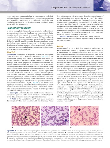
Erythrocytes Anisocytosis is the earliest recognizable morphologic stores are depleted before the red cell mass is compromised. Thus, evalua-
change of erythrocytes in iron-deficiency anemia (Fig. 43–2). The tion of iron stores should be a sensitive and usually reliable means for the
123
anisocytosis is typically accompanied by mild ovalocytosis. As the iron differentiation between iron-deficiency anemia and all other anemias.
deficiency worsens, a mild normochromic, normocytic anemia often Decreased or absent hemosiderin in the marrow is characteristic of iron
develops. With further progression, hemoglobin concentration, ery- deficiency, and is readily evaluated after staining by the simple Prussian
throcyte count, mean corpuscular volume (MCV), and mean erythro- blue method. Stored iron in the macrophages of the marrow can be seen
cyte hemoglobin content all decline together. 124,125 As the indices change in marrow spicules in marrow sections, or in marrow aspirate films. Iron
the erythrocytes appear microcytic and hypochromic on stained blood granules, normally found in the cytoplasm of approximately 30 percent
films. Target cells may sometimes be present. Elongated hypochromic of erythroblasts, become rare but may not be entirely absent.
elliptocytes may be seen, in which the long sides are nearly parallel. Evaluation of the amount of iron in marrow macrophages has long
Such cells have been called “pencil cells,” although they more nearly been considered the “gold standard” for the diagnosis of iron deficiency.
resemble cigars in shape. The red cell indices are consistently abnormal There are, however, technical barriers to the accurate histochemical
in adults only when iron-deficiency anemia is moderate or severe (e.g., determination of marrow iron. First, an invasive procedure, marrow
in males with hemoglobin concentrations <12 g/dL or in women with aspiration, is required. Second, the differentiation of iron within mac-
hemoglobin concentrations <10 g/dL) (Fig. 43–3). The distribution of rophages from artifacts takes experience and skill. In one study only
erythrocyte volume (e.g., red cell distribution width [RDW]) is usually 74 of 108 cases had been accurately reported. Moreover, misleading
131
increased in established iron-deficiency anemia. The RDW is reported results may be obtained in patients who have been transfused or who
often as the coefficient of variation (in percent) of erythrocyte volume have been treated with parenteral iron. The marrow of such patients
132
(see “Differential Diagnosis” below). may contain normal, or even increased, quantities of stainable iron
Leukocytes Leukopenia has been found in some patients with in the face of typical iron-responsive iron-deficiency anemia. In such
iron-deficiency anemia, but the overall distribution of leukocyte counts patients, iron that is seen on marrow examination is not readily avail-
in iron-deficient patients seems to be approximately normal. able for erythropoiesis. As serum markers of iron deficiency became
Platelets Both thrombocytopenia and, more commonly, widely available, the reasons for the primacy of marrow iron estimation
126
thrombocytosis have been associated with iron deficiency. Platelet have been questioned. 133
127
A B C
Figure 43–2. Variability in morphologic diagnosis of iron-deficiency anemia from blood film. As in all deficiency states leading to anemia, the blood
film morphology and blood cell changes are a function of the severity of the deficiency. A. Normal blood film. Normocytic-normochromic red cells
with normal shape. B. Mild iron deficiency. Serum iron, ferritin, and transferring saturation were consistent with mild iron deficiency. Cannot discern if
mean red cell size has decreased. There may be a few red cells that have larger central pallor, but that is arguable. A few cells have oval or elliptical shape.
C. Severe iron deficiency. Serum iron, ferritin, and transferring saturation were consistent with severe iron deficiency. Note obvious increase in overtly
hypochromic cells and higher frequency of microcytes. (Reproduced with permission from Lichtman’s Atlas of Hematology, www.accessmedicine.com.)
Kaushansky_chapter 43_p0627-0650.indd 633 9/17/15 6:27 PM