
Page 648 - Williams Hematology ( PDFDrive )
P. 648
622 Part VI: The Erythrocyte Chapter 42: Iron Metabolism 623
Tf Fe 3+ Tf INTRACELLULAR IRON HOMEOSTASIS
2–
Each cell must regulate its iron uptake and subcellular distribution,
both to assure adequate iron for a multitude of cellular enzymes and
Apo-Tf to prevent excessive iron accumulation that could be injurious or deny
adequate iron to other cells. Accordingly, the synthesis of key cellular
proteins involved in iron transport, storage, and use is regulated post-
Acidification transcriptionally by cellular iron concentrations. 82,83 The mRNA for
STEAP3 TfR1 each of these proteins contains one or several IREs. If the IRE is located
at the 5′ end of the mRNA, it serves to regulate translation; 3′ IREs
Reduction regulate the stability of the mRNA. Each IRE consists of a stem and
NAD(P)H
NAD
H + loop structure, in which the loop is the nucleotide sequence CAGUG
H + 1e Fe 2+ (Fig. 42–7). IRE/IRP–regulated mRNAs include those encoding fer-
Fe 3+ DMT-1 ritin, TfR1, aminolevulinic acid (ALA) synthase, transferrin, aconitase,
DMT-1, and ferroportin. The ferritin mRNA has, as its IRE, a single
Fe 2+ stem–loop structure in the 5′ (upstream) region. In contrast to the fer-
ritin IRE, there are as many as five stems–loops in the 3′ untranslated
portion of TfR mRNA. The IREs are targeted by specific RNA-binding
proteins, IRPs. IRP-1 is cytoplasmic aconitase with four iron-sulfur
clusters and the ability to bind iron, which is required for its aconitase
activity; IRP-2 is highly homologous to IRP-1 but differs by the presence of
3+
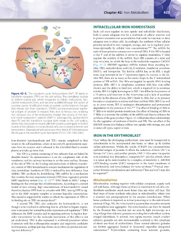
Figure 42–5. The transferrin cycle. Holotransferrin (Fe -Tf) binds to a 73-amino-acid insertion in the N-terminus and a lack of aconitase
2
transferrin receptors (TfR1) on the cell surface. The complexes localize activity. In the absence of iron, IRP-1 binds to IREs, but in its presence
to clathrin-coated pits, which invaginate to initiate endocytosis. Spe- becomes a cytoplasmic aconitase and does not bind IREs. IRP-2 (as well
cialized endosomes form, and become acidified through the action of
a proton pump. Acidification leads to protein conformational changes as, to some extent, IRP-1) undergoes ubiquitination and proteasomal
that release iron from transferrin. STEAP3 (six-transmembrane epithe- degradation in the presence of iron. 86,87 The effect of binding of IRPs to
lial antigen of prostate 3) reduces ferric iron to ferrous iron, enabling 5′ IREs is to inhibit protein translation; the effect of binding of IRPs to
iron transport out of the endosomes through the activity of the diva- 3′ IREs is to increase the stability of the mRNA and thus to enhance the
lent metal transporter-1 protein (DMT-1). Subsequently, apotransferrin synthesis of the gene product. Figure 42–6 illustrates these relationships
(Apo-Tf) and the transferrin receptor both return to the cell surface, for the regulation of synthesis of ferritin and TfR. The net effect of the
where they dissociate at neutral pH. Both proteins participate in further IRE/IRP system is to balance cellular iron uptake with storage, use, and
rounds of iron delivery. In nonerythroid cells, iron is stored as ferritin and in some cell types, export of iron.
hemosiderin. (Reproduced with permission from McKie AT: A ferrireductase
fills the gap in the transferrin cycle. Nat Genet 37(11):1159–1160, 2005.)
IRON IN THE ERYTHROBLAST
Iron-depleted (apo)transferrin and TfR1 remain complexed as they Once within the developing erythroblast, iron must be transported to
return to the cell membrane, where at neutral pH, apotransferrin sepa- mitochondria to be incorporated into heme, or taken up by ferritin
rates from its receptor and is released to the interstitial fluid to reenter within siderosomes. Within the vesicle, STEAP3 (six-transmembrane
3+
plasma and take up more iron. epithelial antigen of prostate 3) effects the reduction of ferric (Fe ) to
2+
The TfR is a protein consisting of two subunits that are linked by ferrous (Fe ) iron, and another protein, DMT-1 (the same transporter
2+
disulfide bonds. Its aminoterminus is on the cytoplasmic side of the as in intestinal iron absorption), transports Fe into the cytosol, where
9
membrane, and its carboxyl-terminus is on the outer surface. Because it is taken up by mitochondria by a complex of mitoferrin-1, ABCB10
of the role of TfR1 in the binding and endocytosis of diferric transfer- (ATP-binding cassette [ABC] transporter in the inner membrane of
88
rin, control of TfR1 biosynthesis is a major mechanism for regulation of mitochondria) and ferrochelatase for heme synthesis. Physical inter-
iron metabolism. Synthesis of TfR1 is induced by iron deficiency. Iron action between mitochondria and endosomes (“kiss and run”) may also
inhibits TfR1 synthesis by destabilizing TfR1 mRNA by a mechanism be required. 89
that involves the iron-responsive element (IRE)/iron-regulatory protein
(IRP) regulatory system (Fig. 42–6). 82,83 TFR1 binds to HFE, using a Mitochondrial Iron
61
binding site that overlaps that of holotransferrin. According to a current Mitochondria, working together with cellular cytoplasm, supply each
model of iron sensing, high concentrations of holotransferrin would cell with heme. Although heme synthesis is important for all cells, ery-
therefore displace HFE from its complex with TfR1, leaving HFE to sig- throblasts synthesize much more heme than any other cell type. The
nal to the BMP receptor complex to increase hepcidin transcription. final steps of heme synthesis take place in mitochondria, where iron
This model is supported by studies in which the expression of HFE or is inserted into protoporphyrin by the enzyme ferrochelatase. When
its binding site on TfR1 are manipulated. 61 heme synthesis is impaired, as in lead poisoning or in the sideroblastic
A second TfR, TfR2, also endocytic for holotransferrin, is not anemias (Chap. 59), the mitochondria accumulates excessive amounts
thought to be involved in delivering iron to cells but its hepatic expres- of amorphous iron aggregates. The mitochondria can then be stained
sion is necessary for normal hepcidin expression and regulation. TfR2 by the Prussian blue reaction and are seen by light microscopy as a
84
influences the BMP complex and its signaling pathway to regulate hep- ring of large blue siderotic granules encircling the erythroblast nucleus
cidin transcription but the molecular mechanism of this effect is not (ringed sideroblast). In normal, iron-replete marrow, (much smaller)
yet understood. TfR2 is also expressed in erythroid precursors where siderotic granules are also demonstrable, scattered in the cytoplasm
it interacts with the erythropoietin receptor and negatively modulates of about one-third of erythroblasts. These normal siderotic granules
erythropoiesis, perhaps putting a brake on erythrocyte production dur- are ferritin aggregates located in lysosomal organelles designated
ing iron deficiency. 85 siderosomes. Erythroblasts containing these siderotic granules,
90
Kaushansky_chapter 42_p0617-0626.indd 623 9/17/15 6:26 PM