
Page 694 - Williams Hematology ( PDFDrive )
P. 694
668 Part VI: The Erythrocyte Chapter 46: Erythrocyte Membrane Disorders 669
Na-K
pump Na-H
Na-K-2Cl Exchange
Cotransporter
3Na 2K
H Na
2CI Na-K
CI/HCO 3 H O
2
Anion exchanger Aquaporin 1
CI/HCO 3 band 3 H O
2
CI K
K H Ca
K-CI
Cotransporter
Gardos Ca Pump
channel
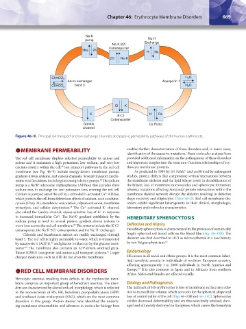
Figure 46–9. Principal ion transport and ion exchange channels and passive permeability pathways of the human erythrocyte.
MEMBRANE PERMEABILITY enabled further characterization of these disorders and, in many cases,
identification of the causative mutations. These molecular analyses have
The red cell membrane displays selective permeability to cations and provided additional information on the pathogenesis of these disorders
anions and it maintains a high potassium, low sodium, and very low and important insights into the structure–function relationships of ery-
calcium content within the cell. Ion transport pathways in the red cell throcyte membrane proteins.
58
membrane (see Fig. 46–9) include energy-driven membrane pumps, As predicted in 1984 by Jiri Palek and confirmed by subsequent
61
gradient-driven systems, and various channels. Several transport mecha- studies, protein defects that compromise vertical interactions between
nisms exist for cations, including two energy-driven pumps. The sodium the membrane skeleton and the lipid bilayer result in destabilization of
58
pump is a Na K adenosine triphosphatase (ATPase) that extrudes three the bilayer, loss of membrane microvesicles and spherocyte formation;
+
+
sodium ions in exchange for two potassium ions entering the red cell. whereas mutations affecting horizontal protein interactions within the
Calcium is pumped out of the cell by a calmodulin-activated Ca ATPase, membrane skeletal network disrupt the skeleton resulting in defective
2+
which protects the cell from deleterious effects of calcium, such as echino- shape recovery and elliptocytes (Table 46–2). Red cell membrane dis-
cytosis (Chap. 31), membrane vesiculation, calpain activation, membrane orders exhibit significant heterogeneity in their clinical, morphologic,
proteolysis, and cellular dehydration. The Ca -activated K channel, laboratory and molecular characteristics.
+
2+
58
also called the Gardos channel, causes selective loss of K in response
+
to increased intracellular Ca . The Na K gradient established by the HEREDITARY SPHEROCYTOSIS
+
+
2+
sodium pump is used by several passive, gradient-driven systems to
move ions across the red cell membrane. The systems include the K Cl Definition and History
+
–
58
cotransporter, the Na K 2Cl cotransporter, and the Na H exchanger. Hereditary spherocytosis is characterized by the presence of osmotically
+
+
+
+
–
Chloride and bicarbonate anions are readily exchanged through fragile spherical red blood cells on the blood film (Fig. 46–10B). The
band 3. The red cell is highly permeable to water, which is transported disorder was first described in 1871 as microcythemia in a case history
by aquaporin-1 (AQP1), and glucose is taken up by the glucose trans- by two Belgian physicians. 62
59
porter. The membrane also contains an ATP-driven oxidized gluta-
60
thione (GSSG) transporter and amino acid transport systems. Larger Epidemiology
58
charged molecules, such as ATP, do not cross the membrane. HS occurs in all racial and ethnic groups. It is the most common inher-
ited hemolytic anemia in individuals of northern European ancestry,
affecting approximately 1 in 2000 individuals in North America and
63
RED CELL MEMBRANE DISORDERS Europe. It is also common in Japan and in Africans from southern
Africa. Males and females are affected equally.
Hemolytic anemias resulting from defects in the erythrocyte mem-
brane comprise an important group of hereditary anemias. The disor- Etiology and Pathogenesis
ders are characterized by altered red cell morphology, which is reflected The hallmark of HS erythrocytes is loss of membrane surface area rela-
in the nomenclature of HS, HE, hereditary pyropoikilocytosis (HPP) tive to intracellular volume, which accounts for the spherical shape and
and southeast Asian ovalocytosis (SAO), which are the most common loss of central pallor of the cell (Figs 46–10B and 46–11C). Spherocytes
disorders in this group. Protein studies have identified the underly- exhibit decreased deformability and are thus selectively retained, dam-
ing membrane abnormalities and advances in molecular biology have aged and ultimately destroyed in the spleen, which causes the hemolysis
Kaushansky_chapter 46_p0661-0688.indd 669 9/17/15 6:42 PM